

Anti-PD-1 [5C4.B8 (Nivolumab)]
Ab00791-1.1
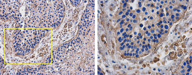
ApplicationsFlow Cytometry, ImmunoHistoChemistry, Neutralisation/Blocking, Other Application
Product group Antibodies
ReactivityHuman, Monkey
TargetPDCD1
Overview
- SupplierAbsolute Antibody
- Product NameAnti-PD-1 [5C4.B8 (Nivolumab)]
- Delivery Days Customer7
- Application Supplier NoteNivolumab has been shown to bind to PD-1-expressing CHO cells (EC50 ~1.66nM). Nivolumab binds CD4+ T cells (EC50 ~0.64 nM) and stains only memory and effector, and not naiive CD4+ or CD8+ T cells from human peripheral blood by FC. The antibody is able to block the interaction between PD-1 and its ligands PDL-1 and PDL-2 (IC50 ~2.52 nM and ~2.59 nM, respectively - determined by SPR) - these IC50 values are also similar to that measured by FACS to evaluate ligand binding to PD-1 expressed on CHO cells. In an allogenic T-cell/DC MLR, Nivolumab-mediated inhibition of PD-1 results in enhancement of IFNgamma release, and also enhances IL-2 secretion (97-139% over an isotype control) in response to the superantigen SEB using human peripheral blood mononuclear cells. The same is also observed in a CMV-restimulation assay. Nivolumab at very low concentrations (~1.5 ng/mL) is able to enhance T-cell reactivity in the presence of a T-cell receptor stimulus - nivolumab has no stimulatory effect in the absence of antigen or T-cell receptor stimulus. In the therapeutically used human IgG4 (S228P) format, this antibody is unable to mediate ADCC (antibody-dependent cell-mediated cytotoxicity) or CDC (complement-dependent cytotoxicity).
- ApplicationsFlow Cytometry, ImmunoHistoChemistry, Neutralisation/Blocking, Other Application
- Applications SupplierIHC; FC; SPR; block
- CertificationResearch Use Only
- ClonalityMonoclonal
- Clone ID5C4.B8 (Nivolumab)
- Gene ID5133
- Target namePDCD1
- Target descriptionprogrammed cell death 1
- Target synonymsADMIO4, AIMTBS, CD279, PD-1, PD1, SLEB2, hPD-1, hPD-l, hSLE1, programmed cell death protein 1, programmed cell death 1 protein, protein PD-1, systemic lupus erythematosus susceptibility 2
- HostMouse
- IsotypeIgG1
- Protein IDQ15116
- Protein NameProgrammed cell death protein 1
- Scientific DescriptionThis chimeric mouse antibody was made using the variable domain sequences of the original human IgG4 (S228P) format, for improved compatibility with existing reagents, assays and techniques. NOT FOR THERAPEUTIC USE- This is a research-grade biosimilar. Th
- ReactivityHuman, Monkey
- Reactivity SupplierHuman, cynomolgus
- Reactivity Supplier NotemAb PD1.5 was prepared by immunizing IgH and IgK knock-out transgenic mice possessing a human immunoglobulin (heavy chain) minilocus with recombinant human PD-1-Fc protein consisting of the extracellular domain of PD-1 (amino acids 1–167) and the Fc portion of human IgG1, and Chinese hamster ovary (CHO) cells expressing human PD-1. Nivolumab was generated by grafting the variable regions of PD1.5 onto human kappa and IgG4 constant regions containing an S228P mutation (prevents Fab arm exchange with endogenous IgG4 antibodies).
- Storage Instruction-20°C,2°C to 8°C
- UNSPSC41116161
Related products
Product group Antibodies
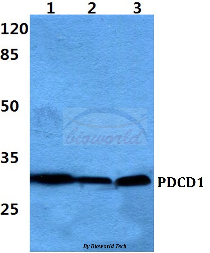
Anti-PDCD1 AntibodyA28569
ApplicationsWestern Blot
ReactivityHuman, Mouse, Rat
- SizePrice
Product group Antibodies
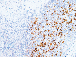
anti-PD-1 (human), mAb (AG-IHC001)AG-20B-6020
ApplicationsELISA, ImmunoHistoChemistry
ReactivityHuman
TargetPDCD1
- SizePrice

Product group Antibodies
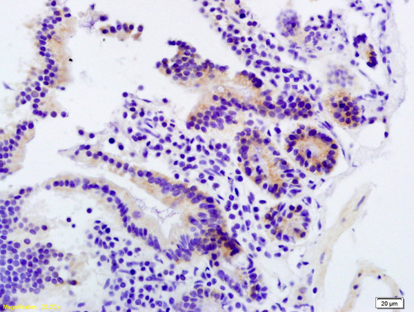
Anti-PDCD1 AntibodyAMAB91197
ApplicationsWestern Blot, ImmunoHistoChemistry
ReactivityHuman
TargetPDCD1
- SizePrice
Product group Antibodies

ApplicationsFlow Cytometry, ImmunoHistoChemistry
ReactivityMouse
TargetPDCD1
- SizePrice
Product group Antibodies
References
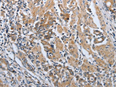
PD-1 Polyclonal AntibodyBS-1867R
ApplicationsFlow Cytometry, ImmunoFluorescence, Western Blot, ELISA, ImmunoCytoChemistry, ImmunoHistoChemistry, ImmunoHistoChemistry Frozen, ImmunoHistoChemistry Paraffin
ReactivityHuman, Mouse, Rat
TargetPDCD1
- SizePrice
Product group Antibodies
PDCD1 AntibodyCSB-PA483633
ApplicationsELISA, ImmunoHistoChemistry
ReactivityHuman
TargetPDCD1
- SizePrice
Product group Antibodies
Pdcd1 Polyclonal AntibodyCAC07144
ApplicationsImmunoFluorescence, Western Blot, ELISA, ImmunoHistoChemistry
ReactivityMouse
TargetPDCD1
- SizePrice
Product group Antibodies
PD1 antibodyGTX128435
ApplicationsWestern Blot, ImmunoHistoChemistry, ImmunoHistoChemistry Paraffin
ReactivityHuman, Mouse
TargetPDCD1
- SizePrice