

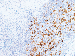
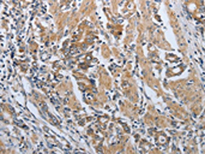
Formalin-fixed and paraffin embedded mouse colon labeled with Anti-PD-1/CD279 Polyclonal Antibody, Unconjugated (bs-1867R) at 1:200 followed by conjugation to the secondary antibody and DAB staining.
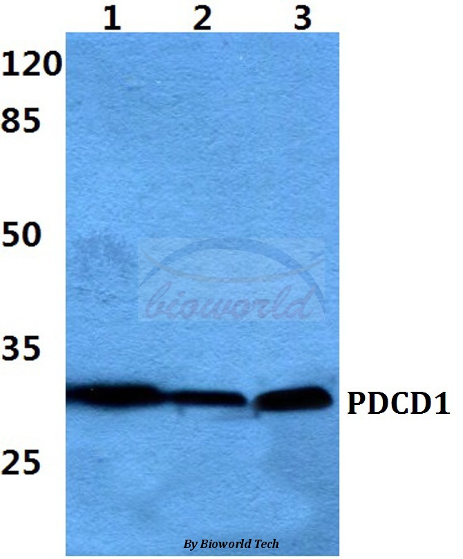
 at 1:200 overnight at 4˚C. Followed by conjugation to secondary antibody (bs-0295G-HRP) at 1:3000 for 90 min at 37˚C. Predicted band 30kD. Observed band size:30kD.\n")
 at 1:500 overnight at 4°C followed by a conjugated secondary antibody for 60 minutes at 37°C.")
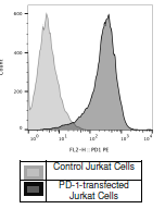
 (green) at 1:100 for 30 minutes at room temperature. The graph compares PD-1 antibody to unstained cells (blue) and isotype control (orange).")
 at 1:1000 dilution and 4˚C overnight incubation. Followed by conjugated secondary antibody incubation at 1:20000 for 60 min at 37˚C.")
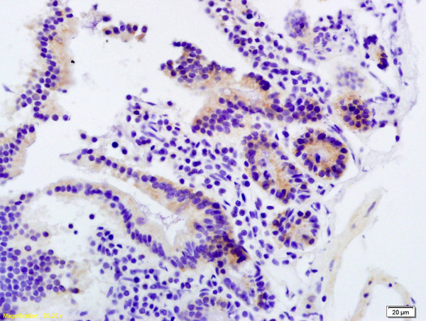
; Antigen retrieval by boiling in sodium citrate buffer (pH6.0) for 15min; Block endogenous peroxidase by 3% hydrogen peroxide for 20 minutes; Blocking buffer (normal goat serum) at 37°C for 30min; Antibody incubation with (PD-1 ) Polyclonal Antibody, Unconjugated (bs-1867R) at 1:200 overnight at 4°C, followed by operating according to SP Kit(Rabbit) (sp-0023) instructionsand DAB staining.")
; Antigen retrieval by boiling in sodium citrate buffer (pH6.0) for 15min; Block endogenous peroxidase by 3% hydrogen peroxide for 20 minutes; Blocking buffer (normal goat serum) at 37°C for 30min; Antibody incubation with (PD-1 ) Polyclonal Antibody, Unconjugated (bs-1867R) at 1:200 overnight at 4°C, followed by operating according to SP Kit(Rabbit) (sp-0023) instructionsand DAB staining.")
Formalin-fixed and paraffin embedded mouse colon labeled with Anti-PD-1/CD279 Polyclonal Antibody, Unconjugated (bs-1867R) at 1:200 followed by conjugation to the secondary antibody and DAB staining.
PD-1 Polyclonal Antibody
BS-1867R
ApplicationsFlow Cytometry, ImmunoFluorescence, Western Blot, ELISA, ImmunoCytoChemistry, ImmunoHistoChemistry, ImmunoHistoChemistry Frozen, ImmunoHistoChemistry Paraffin
Product group Antibodies
ReactivityHuman, Mouse, Rat
TargetPDCD1
Overview
- SupplierBioss
- Product NamePD-1 Polyclonal Antibody
- Delivery Days Customer16
- ApplicationsFlow Cytometry, ImmunoFluorescence, Western Blot, ELISA, ImmunoCytoChemistry, ImmunoHistoChemistry, ImmunoHistoChemistry Frozen, ImmunoHistoChemistry Paraffin
- Applications SupplierWB(1:300-5000), ELISA(1:500-1000), FCM(1:20-100), IHC-P(1:200-400), IHC-F(1:100-500), IF(IHC-P)(1:50-200), IF(IHC-F)(1:50-200), IF(ICC)(1:50-200)
- CertificationResearch Use Only
- ClonalityPolyclonal
- Concentration1 ug/ul
- ConjugateUnconjugated
- Gene ID5133
- Target namePDCD1
- Target descriptionprogrammed cell death 1
- Target synonymsADMIO4, AIMTBS, CD279, PD-1, PD1, SLEB2, hPD-1, hPD-l, hSLE1, programmed cell death protein 1, programmed cell death 1 protein, protein PD-1, systemic lupus erythematosus susceptibility 2
- HostRabbit
- IsotypeIgG
- Protein IDQ15116
- Protein NameProgrammed cell death protein 1
- ReactivityHuman, Mouse, Rat
- Storage Instruction-20°C
- UNSPSC41116161
References
- Programmed cell death protein 1 promotes hepatitis B virus transmission through the regulation of ERK1/2-mediated trophoblasts differentiation. Yang J et al., 2020 Feb, Arch Gynecol ObstetRead this paper
- Expansion of circulating follicular T helper cells associates with disease severity in childhood atopic dermatitis. Szabo K et al., 2017 Sep, Immunol LettRead this paper
- Effect of EBI3 on radiation-induced immunosuppression of cervical cancer HeLa cells by regulating Treg cells through PD-1/PD-L1 pathway. Zhang SA et al., 2017 Mar, Tumour BiolRead this paper
Datasheet
Related products
Product group Antibodies
Anti-PDCD1 AntibodyA28569
ApplicationsWestern Blot
ReactivityHuman, Mouse, Rat
- SizePrice
Product group Antibodies
anti-PD-1 (human), mAb (AG-IHC001)AG-20B-6020
ApplicationsELISA, ImmunoHistoChemistry
ReactivityHuman
TargetPDCD1
- SizePrice
Product group Antibodies
ApplicationsNeutralisation/Blocking
TargetPDCD1
- SizePrice

Product group Antibodies
Anti-PDCD1 AntibodyAMAB91197
ApplicationsWestern Blot, ImmunoHistoChemistry
ReactivityHuman
TargetPDCD1
- SizePrice
Product group Antibodies

ApplicationsFlow Cytometry, ImmunoHistoChemistry
ReactivityMouse
TargetPDCD1
- SizePrice
Product group Antibodies

Anti-PD-1 [5C4.B8 (Nivolumab)]Ab00791-1.1
ApplicationsFlow Cytometry, ImmunoHistoChemistry, Neutralisation/Blocking, Other Application
ReactivityHuman, Monkey
TargetPDCD1
- SizePrice
Product group Antibodies
PDCD1 AntibodyCSB-PA483633
ApplicationsELISA, ImmunoHistoChemistry
ReactivityHuman
TargetPDCD1
- SizePrice
Product group Antibodies
Pdcd1 Polyclonal AntibodyCAC07144
ApplicationsImmunoFluorescence, Western Blot, ELISA, ImmunoHistoChemistry
ReactivityMouse
TargetPDCD1
- SizePrice