


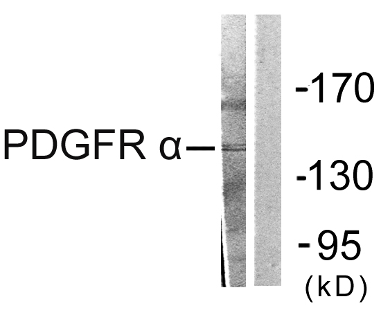
Figure 1. Western blot analysis of PDGFRA using anti-PDGFRA antibody (A00366). Electrophoresis was performed on a 5-20% SDS-PAGE gel at 70V (Stacking gel) / 90V (Resolving gel) for 2-3 hours. The sample well of each lane was loaded with 30 ug of sample under reducing conditions. Lane 1: human 293T whole cell lysates, Lane 2: human U251 whole cell lysates, Lane 3: rat brain tissue lysates, Lane 4: rat C6 whole cell lysates, Lane 5: mouse brain tissue lysates, Lane 6: mouse NIH/3T3 whole cell lysates. After electrophoresis, proteins were transferred to a nitrocellulose membrane at 150 mA for 50-90 minutes. Blocked the membrane with 5% non-fat milk/TBS for 1.5 hour at RT. The membrane was incubated with rabbit anti-PDGFRA antigen affinity purified polyclonal antibody (Catalog # A00366) at 0.5 microg/mL overnight at 4°C, then washed with TBS-0.1%Tween 3 times with 5 minutes each and probed with a goat anti-rabbit IgG-HRP secondary antibody at a dilution of 1:5000 for 1.5 hour at RT. The signal is developed using an Enhanced Chemiluminescent detection (ECL) kit (Catalog # EK1002) with Tanon 5200 system. A specific band was detected for PDGFRA at approximately 180 kDa. The expected band size for PDGFRA is at 123,180 kDa.
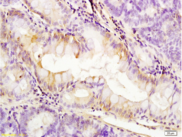
. PDGFRA was detected in an immunocytochemical section of CACO2 cells. Enzyme antigen retrieval was performed using IHC enzyme antigen retrieval reagent (AR0022) for 15 mins. The cells were blocked with 10% goat serum. And then incubated with 5 microg/mL rabbit anti-PDGFRA Antibody (A00366) overnight at 4°C. Cy3 Conjugated Goat Anti-Rabbit IgG (BA1032) was used as secondary antibody at 1:500 dilution and incubated for 30 minutes at 37°C. The section was counterstained with DAPI. Visualize using a fluorescence microscope and filter sets appropriate for the label used.")
Figure 1. Western blot analysis of PDGFRA using anti-PDGFRA antibody (A00366). Electrophoresis was performed on a 5-20% SDS-PAGE gel at 70V (Stacking gel) / 90V (Resolving gel) for 2-3 hours. The sample well of each lane was loaded with 30 ug of sample under reducing conditions. Lane 1: human 293T whole cell lysates, Lane 2: human U251 whole cell lysates, Lane 3: rat brain tissue lysates, Lane 4: rat C6 whole cell lysates, Lane 5: mouse brain tissue lysates, Lane 6: mouse NIH/3T3 whole cell lysates. After electrophoresis, proteins were transferred to a nitrocellulose membrane at 150 mA for 50-90 minutes. Blocked the membrane with 5% non-fat milk/TBS for 1.5 hour at RT. The membrane was incubated with rabbit anti-PDGFRA antigen affinity purified polyclonal antibody (Catalog # A00366) at 0.5 microg/mL overnight at 4°C, then washed with TBS-0.1%Tween 3 times with 5 minutes each and probed with a goat anti-rabbit IgG-HRP secondary antibody at a dilution of 1:5000 for 1.5 hour at RT. The signal is developed using an Enhanced Chemiluminescent detection (ECL) kit (Catalog # EK1002) with Tanon 5200 system. A specific band was detected for PDGFRA at approximately 180 kDa. The expected band size for PDGFRA is at 123,180 kDa.
Anti-PDGFRA Antibody Picoband(r)
A00366-CARRIER-FREE
ApplicationsImmunoFluorescence, Western Blot, ELISA, ImmunoCytoChemistry
Product group Antibodies
ReactivityHuman, Mouse, Rat
TargetPDGFRA
Overview
- SupplierBoster Bio
- Product NameAnti-PDGFRA Antibody Picoband(r)
- Delivery Days Customer9
- Application Supplier NoteTested Species: In-house tested species with positive results. Other applications have not been tested. Optimal dilutions should be determined by end users.
- ApplicationsImmunoFluorescence, Western Blot, ELISA, ImmunoCytoChemistry
- CertificationResearch Use Only
- ClonalityPolyclonal
- Concentration500 ug/ml
- Gene ID5156
- Target namePDGFRA
- Target descriptionplatelet derived growth factor receptor alpha
- Target synonymsCD140A, PDGFR-2, PDGFR2, platelet-derived growth factor receptor alpha, CD140 antigen-like family member A, CD140a antigen, PDGF-R-alpha, alpha-type platelet-derived growth factor receptor, platelet-derived growth factor receptor 2, platelet-derived growth factor receptor, alpha polypeptide
- HostRabbit
- IsotypeIgG
- Protein IDP16234
- Protein NamePlatelet-derived growth factor receptor alpha
- Scientific DescriptionBoster Bio Anti-PDGFRA Antibody Picoband® catalog # A00366. Tested in ELISA, IF, ICC, WB applications. This antibody reacts with Human, Mouse, Rat. The brand Picoband indicates this is a premium antibody that guarantees superior quality, high affinity, and strong signals with minimal background in Western blot applications. Only our best-performing antibodies are designated as Picoband, ensuring unmatched performance.
- ReactivityHuman, Mouse, Rat
- Storage Instruction-20°C,2°C to 8°C
- UNSPSC12352203
Related products
Product group Antibodies
ApplicationsImmunoFluorescence, Western Blot, ELISA, ImmunoHistoChemistry
ReactivityHuman, Mouse, Rat
- SizePrice
Product group Antibodies
Anti-PDGFRA Antibody144-02103
ApplicationsWestern Blot
ReactivityHuman, Mouse, Rat
TargetPDGFRA
- SizePrice
Product group Antibodies

ApplicationsImmunoFluorescence, ImmunoHistoChemistry, ImmunoHistoChemistry Paraffin
ReactivityHuman, Mouse, Rat
TargetPDGFRA
- SizePrice
Product group Antibodies
References
PDGFRA Polyclonal AntibodyBS-0231R
ApplicationsImmunoFluorescence, ELISA, ImmunoCytoChemistry, ImmunoHistoChemistry, ImmunoHistoChemistry Frozen, ImmunoHistoChemistry Paraffin
ReactivityBovine, Canine, Chicken, Equine, Human, Mouse, Porcine, Rat
TargetPDGFRA
- SizePrice
Product group Antibodies
PDGFRA AntibodyCSB-PA003724
ApplicationsImmunoFluorescence, Western Blot, ELISA, ImmunoHistoChemistry
ReactivityHuman, Mouse, Rat
TargetPDGFRA
- SizePrice
Product group Antibodies
Pdgfra Polyclonal AntibodyCAC08107
ApplicationsImmunoFluorescence, Western Blot, ELISA
ReactivityMouse
TargetPDGFRA
- SizePrice

Product group Antibodies
Anti-PDGFRA AntibodyHPA004947
ApplicationsImmunoCytoChemistry
ReactivityHuman
TargetPDGFRA
- SizePrice
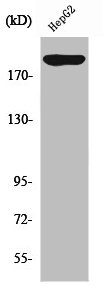
![Human tissue extract (30 μg) was separated by 5% SDS-PAGE, and the membrane was blotted with PDGF Receptor alpha antibody [N2C2], Internal (GTX107903) diluted at 1:1000. The HRP-conjugated anti-rabbit IgG antibody (GTX213110-01) was used to detect the primary antibody, and the signal was developed with Trident ECL plus-Enhanced.](https://www.genetex.com/upload/website/prouct_img/normal/GTX107903/GTX107903_39988_20220617_WB_skeletalmuscle_22062121_572.webp)
Product group Antibodies
ApplicationsImmunoFluorescence, Western Blot, ImmunoCytoChemistry
ReactivityHuman, Mouse, Rat
TargetPDGFRA
- SizePrice