

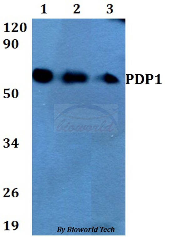
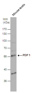
Anti-PDP1 Antibody
A28851
ApplicationsWestern Blot
Product group Antibodies
ReactivityHuman, Mouse
Overview
- SupplierAntibodies.com
- Product NameAnti-PDP1 Antibody
- Delivery Days Customer7
- ApplicationsWestern Blot
- CertificationResearch Use Only
- ClonalityPolyclonal
- ConjugateUnconjugated
- Estimated Purity>95%
- HostRabbit
- Scientific DescriptionRabbit polyclonal antibody to PDP1
- ReactivityHuman, Mouse
- UNSPSC12352203
Related products

Product group Antibodies
Anti-PDP1/PDP Antibody Picoband(r)A05607-2-CARRIER-FREE
ApplicationsFlow Cytometry, ImmunoFluorescence, Western Blot, ELISA, ImmunoCytoChemistry
ReactivityHuman, Mouse, Rat
TargetPDP1
- SizePrice
Product group Antibodies
Anti-PDP1 Antibody144-60701
ApplicationsImmunoFluorescence, Western Blot
ReactivityHuman, Mouse, Rat
TargetPDP1
- SizePrice
Product group Antibodies
PPM2C Polyclonal AntibodyBS-3966R
ApplicationsImmunoFluorescence, Western Blot, ELISA, ImmunoCytoChemistry, ImmunoHistoChemistry, ImmunoHistoChemistry Frozen, ImmunoHistoChemistry Paraffin
ReactivityBovine, Canine, Equine, Human, Mouse, Rabbit, Rat
TargetPDP1
- SizePrice
Product group Antibodies
PDP1 AntibodyCSB-PA113942
ApplicationsELISA, ImmunoHistoChemistry
ReactivityHuman, Mouse, Rat
TargetPDP1
- SizePrice
Product group Antibodies
PDP1 Polyclonal AntibodyCAC15388
ApplicationsWestern Blot, ELISA, ImmunoHistoChemistry
TargetPDP1
- SizePrice
Product group Antibodies

PDP1 AntibodyLS-C401943
ApplicationsELISA, ImmunoHistoChemistry
ReactivityHuman, Mouse, Rat
TargetPDP1
- SizePrice
Product group Antibodies
PDP1 antibody [N1N3]GTX109533
ApplicationsWestern Blot
ReactivityHuman, Mouse
TargetPDP1
- SizePrice

Product group Antibodies
Anti-PDP1 AntibodyHPA018483
ApplicationsWestern Blot, ImmunoHistoChemistry
ReactivityHuman
TargetPDP1
- SizePrice
Product group Antibodies
Anti-PDP1 AntibodyCAB14545
ApplicationsImmunoFluorescence, Western Blot, ELISA, ImmunoCytoChemistry
ReactivityHuman
TargetPDP1
- SizePrice