

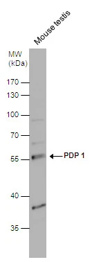
PDP 1 antibody detects PDP 1 protein by western blot analysis. Mouse tissue extracts (50 μg) was separated by 10% SDS-PAGE, and the membrane was blotted with PDP 1 antibody (GTX109533) diluted at 1:1000.
PDP 1 antibody detects PDP 1 protein by western blot analysis. Mouse tissue extracts (50 μg) was separated by 10% SDS-PAGE, and the membrane was blotted with PDP 1 antibody (GTX109533) diluted at 1:1000.
PDP1 antibody [N1N3]
GTX109533
ApplicationsWestern Blot
Product group Antibodies
ReactivityHuman, Mouse
TargetPDP1
Overview
- SupplierGeneTex
- Product NamePDP1 antibody [N1N3]
- Delivery Days Customer9
- Application Supplier NoteWB: 1:500-1:3000. *Optimal dilutions/concentrations should be determined by the researcher.Not tested in other applications.
- ApplicationsWestern Blot
- CertificationResearch Use Only
- ClonalityPolyclonal
- Concentration1 mg/ml
- ConjugateUnconjugated
- Gene ID54704
- Target namePDP1
- Target descriptionpyruvate dehydrogenase phosphatase catalytic subunit 1
- Target synonymsPDH, PDP, PDPC, PDPC 1, PPM2A, PPM2C, pyruvate dehyrogenase phosphatase catalytic subunit 1, [Pyruvate dehydrogenase [acetyl-transferring]]-phosphatase 1, mitochondrial, protein phosphatase 2C, magnesium-dependent, catalytic subunit, protein phosphatase, Mg2+/Mn2+ dependent 2A, pyruvate dehydrogenase (Lipoamide) phosphatase-phosphatase
- HostRabbit
- IsotypeIgG
- Protein IDQ9P0J1
- Protein Name[Pyruvate dehydrogenase [acetyl-transferring]]-phosphatase 1, mitochondrial
- Scientific DescriptionThis gene encodes a protein similar to members of the protein phosphatase 2C (PP2C) family. Studies of the rat counterpart of this protein suggested that this protein may reside within the mitochondrial matrix space and be responsible for dephosphorylation and reactivation of the pyruvate dehydrogenase complex (PDC). [provided by RefSeq]
- ReactivityHuman, Mouse
- Storage Instruction-20°C or -80°C,2°C to 8°C
- UNSPSC41116161
Datasheet
Related products

Product group Antibodies
Anti-PDP1/PDP Antibody Picoband(r)A05607-2-CARRIER-FREE
ApplicationsFlow Cytometry, ImmunoFluorescence, Western Blot, ELISA, ImmunoCytoChemistry
ReactivityHuman, Mouse, Rat
TargetPDP1
- SizePrice
Product group Antibodies
Anti-PDP1 AntibodyA28851
ApplicationsWestern Blot
ReactivityHuman, Mouse
- SizePrice
Product group Antibodies
Anti-PDP1 Antibody144-60701
ApplicationsImmunoFluorescence, Western Blot
ReactivityHuman, Mouse, Rat
TargetPDP1
- SizePrice
Product group Antibodies
PPM2C Polyclonal AntibodyBS-3966R
ApplicationsImmunoFluorescence, Western Blot, ELISA, ImmunoCytoChemistry, ImmunoHistoChemistry, ImmunoHistoChemistry Frozen, ImmunoHistoChemistry Paraffin
ReactivityBovine, Canine, Equine, Human, Mouse, Rabbit, Rat
TargetPDP1
- SizePrice
Product group Antibodies
PDP1 AntibodyCSB-PA113942
ApplicationsELISA, ImmunoHistoChemistry
ReactivityHuman, Mouse, Rat
TargetPDP1
- SizePrice
Product group Antibodies
PDP1 Polyclonal AntibodyCAC15388
ApplicationsWestern Blot, ELISA, ImmunoHistoChemistry
TargetPDP1
- SizePrice
Product group Antibodies

PDP1 AntibodyLS-C401943
ApplicationsELISA, ImmunoHistoChemistry
ReactivityHuman, Mouse, Rat
TargetPDP1
- SizePrice

Product group Antibodies
Anti-PDP1 AntibodyHPA018483
ApplicationsWestern Blot, ImmunoHistoChemistry
ReactivityHuman
TargetPDP1
- SizePrice
Product group Antibodies
Anti-PDP1 AntibodyCAB14545
ApplicationsImmunoFluorescence, Western Blot, ELISA, ImmunoCytoChemistry
ReactivityHuman
TargetPDP1
- SizePrice