

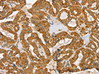
The image on the left is immunohistochemistry of paraffin-embedded Human thyroid cancer tissue using CSB-PA113942(PDP1 Antibody) at dilution 1/30, on the right is treated with fusion protein. (Original magnification: x200)
 at dilution 1/30, on the right is treated with fusion protein. (Original magnification: x200)")
The image on the left is immunohistochemistry of paraffin-embedded Human thyroid cancer tissue using CSB-PA113942(PDP1 Antibody) at dilution 1/30, on the right is treated with fusion protein. (Original magnification: x200)
PDP1 Antibody
CSB-PA113942
ApplicationsELISA, ImmunoHistoChemistry
Product group Antibodies
ReactivityHuman, Mouse, Rat
TargetPDP1
Overview
- SupplierCusabio
- Product NamePDP1 Antibody
- Delivery Days Customer20
- ApplicationsELISA, ImmunoHistoChemistry
- CertificationResearch Use Only
- ClonalityPolyclonal
- ConjugateUnconjugated
- Gene ID54704
- Target namePDP1
- Target descriptionpyruvate dehydrogenase phosphatase catalytic subunit 1
- Target synonymsPDH, PDP, PDPC, PDPC 1, PPM2A, PPM2C, pyruvate dehyrogenase phosphatase catalytic subunit 1, [Pyruvate dehydrogenase [acetyl-transferring]]-phosphatase 1, mitochondrial, protein phosphatase 2C, magnesium-dependent, catalytic subunit, protein phosphatase, Mg2+/Mn2+ dependent 2A, pyruvate dehydrogenase (Lipoamide) phosphatase-phosphatase
- HostRabbit
- IsotypeIgG
- Protein IDQ9P0J1
- Protein Name[Pyruvate dehydrogenase [acetyl-transferring]]-phosphatase 1, mitochondrial
- Scientific DescriptionPyruvate dehydrogenase (E1) is one of the three components (E1, E2, and E3) of the large pyruvate dehydrogenase complex. Pyruvate dehydrogenase kinases catalyze phosphorylation of serine residues of E1 to inactivate the E1 component and inhibit the complex. Pyruvate dehydrogenase phosphatases catalyze the dephosphorylation and activation of the E1 component to reverse the effects of pyruvate dehydrogenase kinases.
- ReactivityHuman, Mouse, Rat
- Storage Instruction-20°C or -80°C
- UNSPSC41116161
Related products

Product group Antibodies
Anti-PDP1/PDP Antibody Picoband(r)A05607-2-CARRIER-FREE
ApplicationsFlow Cytometry, ImmunoFluorescence, Western Blot, ELISA, ImmunoCytoChemistry
ReactivityHuman, Mouse, Rat
TargetPDP1
- SizePrice
Product group Antibodies
Anti-PDP1 AntibodyA28851
ApplicationsWestern Blot
ReactivityHuman, Mouse
- SizePrice
Product group Antibodies
Anti-PDP1 Antibody144-60701
ApplicationsImmunoFluorescence, Western Blot
ReactivityHuman, Mouse, Rat
TargetPDP1
- SizePrice
Product group Antibodies
PPM2C Polyclonal AntibodyBS-3966R
ApplicationsImmunoFluorescence, Western Blot, ELISA, ImmunoCytoChemistry, ImmunoHistoChemistry, ImmunoHistoChemistry Frozen, ImmunoHistoChemistry Paraffin
ReactivityBovine, Canine, Equine, Human, Mouse, Rabbit, Rat
TargetPDP1
- SizePrice
Product group Antibodies
PDP1 Polyclonal AntibodyCAC15388
ApplicationsWestern Blot, ELISA, ImmunoHistoChemistry
TargetPDP1
- SizePrice
Product group Antibodies

PDP1 AntibodyLS-C401943
ApplicationsELISA, ImmunoHistoChemistry
ReactivityHuman, Mouse, Rat
TargetPDP1
- SizePrice
Product group Antibodies
PDP1 antibody [N1N3]GTX109533
ApplicationsWestern Blot
ReactivityHuman, Mouse
TargetPDP1
- SizePrice

Product group Antibodies
Anti-PDP1 AntibodyHPA018483
ApplicationsWestern Blot, ImmunoHistoChemistry
ReactivityHuman
TargetPDP1
- SizePrice
Product group Antibodies
Anti-PDP1 AntibodyCAB14545
ApplicationsImmunoFluorescence, Western Blot, ELISA, ImmunoCytoChemistry
ReactivityHuman
TargetPDP1
- SizePrice