


Figure 1. Western blot analysis of PPARGC1B using anti-PPARGC1B antibody (A02933-1). Electrophoresis was performed on a 5-20% SDS-PAGE gel at 70V (Stacking gel) / 90V (Resolving gel) for 2-3 hours. The sample well of each lane was loaded with 50ug of sample under reducing conditions. Lane 1: human Caco-2 whole cell lysates, Lane 2: human HEK293 whole cell lysates, Lane 3: human U2OS whole cell lysates. Lane 4: human MDA-MB-453 whole cell lysates, Lane 5: human K562 whole cell lysates. After Electrophoresis, proteins were transferred to a Nitrocellulose membrane at 150mA for 50-90 minutes. Blocked the membrane with 5% Non-fat Milk/ TBS for 1.5 hour at RT. The membrane was incubated with rabbit anti-PPARGC1B antigen affinity purified polyclonal antibody (Catalog # A02933-1) at 0.25 microg/mL overnight at 4°C, then washed with TBS-0.1%Tween 3 times with 5 minutes each and probed with a goat anti-rabbit IgG-HRP secondary antibody at a dilution of 1:5000 for 1.5 hour at RT. The signal is developed using an Enhanced Chemiluminescent detection (ECL) kit (Catalog # EK1002) with Tanon 5200 system. A specific band was detected for PPARGC1B at approximately 113KD. The expected band size for PPARGC1B is at 113KD.
. Overlay histogram showing U937 cells stained with A02933-1 (Blue line). To facilitate intracellular staining, cells were fixed with 4% paraformaldehyde and permeabilized with permeabilization buffer. The cells were blocked with 10% normal goat serum. And then incubated with rabbit anti-PPARGC1B Antibody (A02933-1, 1microg/1x106 cells) for 30 min at 20°C. DyLight®488 conjugated goat anti-rabbit IgG (BA1127, 5-10microg/1x106 cells) was used as secondary antibody for 30 minutes at 20°C. Isotype control antibody (Green line) was rabbit IgG (1microg/1x106) used under the same conditions. Unlabelled sample without incubation with primary antibody and secondary antibody (Red line) was used as a blank control.")
. Overlay histogram showing HEPA1-6 cells stained with A02933-1 (Blue line). To facilitate intracellular staining, cells were fixed with 4% paraformaldehyde and permeabilized with permeabilization buffer. The cells were blocked with 10% normal goat serum. And then incubated with rabbit anti-PPARGC1B Antibody (A02933-1, 1microg/1x106 cells) for 30 min at 20°C. DyLight®488 conjugated goat anti-rabbit IgG (BA1127, 5-10microg/1x106 cells) was used as secondary antibody for 30 minutes at 20°C. Isotype control antibody (Green line) was rabbit IgG (1microg/1x106) used under the same conditions. Unlabelled sample without incubation with primary antibody and secondary antibody (Red line) was used as a blank control.")
Figure 1. Western blot analysis of PPARGC1B using anti-PPARGC1B antibody (A02933-1). Electrophoresis was performed on a 5-20% SDS-PAGE gel at 70V (Stacking gel) / 90V (Resolving gel) for 2-3 hours. The sample well of each lane was loaded with 50ug of sample under reducing conditions. Lane 1: human Caco-2 whole cell lysates, Lane 2: human HEK293 whole cell lysates, Lane 3: human U2OS whole cell lysates. Lane 4: human MDA-MB-453 whole cell lysates, Lane 5: human K562 whole cell lysates. After Electrophoresis, proteins were transferred to a Nitrocellulose membrane at 150mA for 50-90 minutes. Blocked the membrane with 5% Non-fat Milk/ TBS for 1.5 hour at RT. The membrane was incubated with rabbit anti-PPARGC1B antigen affinity purified polyclonal antibody (Catalog # A02933-1) at 0.25 microg/mL overnight at 4°C, then washed with TBS-0.1%Tween 3 times with 5 minutes each and probed with a goat anti-rabbit IgG-HRP secondary antibody at a dilution of 1:5000 for 1.5 hour at RT. The signal is developed using an Enhanced Chemiluminescent detection (ECL) kit (Catalog # EK1002) with Tanon 5200 system. A specific band was detected for PPARGC1B at approximately 113KD. The expected band size for PPARGC1B is at 113KD.
Anti-PGC1 beta/PPARGC1B Picoband(r) Antibody
A02933-1-CARRIER-FREE
ApplicationsFlow Cytometry, Western Blot, ELISA
Product group Antibodies
ReactivityHuman, Mouse, Rat
TargetPPARGC1B
Overview
- SupplierBoster Bio
- Product NameAnti-PGC1 beta/PPARGC1B Picoband(r) Antibody
- Delivery Days Customer9
- ApplicationsFlow Cytometry, Western Blot, ELISA
- CertificationResearch Use Only
- ClonalityPolyclonal
- Concentration500 ug/ml
- Gene ID133522
- Target namePPARGC1B
- Target descriptionPPARG coactivator 1 beta
- Target synonymsERRL1, PERC, PGC-1(beta), PGC1B, peroxisome proliferator-activated receptor gamma coactivator 1-beta, PGC-1-related estrogen receptor alpha coactivator, PPAR-gamma coactivator 1-beta, PPARAGCIbeta, PPARGC-1-beta, PPARgamma coactivator 1 beta, peroxisome proliferator-activated receptor gamma, coactivator 1 beta
- HostRabbit
- IsotypeIgG
- Protein IDQ86YN6
- Protein NamePeroxisome proliferator-activated receptor gamma coactivator 1-beta
- Scientific DescriptionBoster Bio Anti-PGC1 beta/PPARGC1B Picoband® Antibody catalog # A02933-1. Tested in ELISA, Flow Cytometry, WB applications. This antibody reacts with Human, Mouse, Rat. The brand Picoband indicates this is a premium antibody that guarantees superior quality, high affinity, and strong signals with minimal background in Western blot applications. Only our best-performing antibodies are designated as Picoband, ensuring unmatched performance.
- ReactivityHuman, Mouse, Rat
- Storage Instruction-20°C,2°C to 8°C
- UNSPSC12352203
Related products
Product group Antibodies
PGC1 beta Recombinant Antibody, Biotin ConjugatedBSM-61701R-BIOTIN
ApplicationsImmunoPrecipitation, Western Blot
ReactivityHuman, Mouse, Rat
TargetPPARGC1B
- SizePrice
Product group Antibodies
Anti-PPARGC1B AntibodyA45009
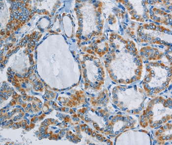
ApplicationsImmunoHistoChemistry
ReactivityHuman
- SizePrice
Product group Antibodies
PPARGC1B AntibodyCSB-PA012670
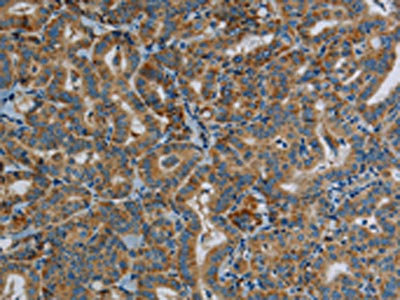
ApplicationsELISA, ImmunoHistoChemistry
ReactivityHuman
TargetPPARGC1B
- SizePrice
Product group Antibodies
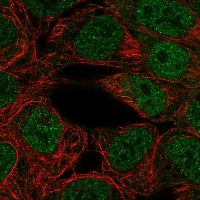
Anti-PPARGC1B AntibodyHPA050543
ApplicationsImmunoCytoChemistry
ReactivityHuman
TargetPPARGC1B
- SizePrice
Product group Antibodies

ApplicationsELISA, ImmunoHistoChemistry
ReactivityCanine, Human, Mouse, Rat
TargetPPARGC1B
- SizePrice
Product group Antibodies
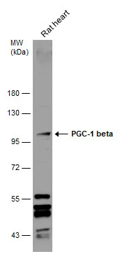
PGC1 beta antibodyGTX129682
ApplicationsWestern Blot
ReactivityHuman, Rat
TargetPPARGC1B
- SizePrice