


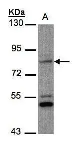
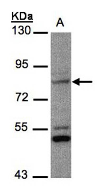
Figure 1. Western blot analysis of PHEX using anti-PHEX antibody (A02078). Electrophoresis was performed on a 5-20% SDS-PAGE gel at 70V (Stacking gel) / 90V (Resolving gel) for 2-3 hours. The sample well of each lane was loaded with 50ug of sample under reducing conditions. Lane 1: rat ovary tissue lysates, Lane 2: rat kidney tissue lysates, Lane 3: mouse ovary tissue lysates, Lane 4: mouse kidney tissue lysates, Lane 5: human HEK293 whole cell lysates, Lane 6: monkey kidney tissue lysates, Lane 7: rat NRK whole cell lysates, Lane 8: monkey COS-7 whole cell lysates. After Electrophoresis, proteins were transferred to a Nitrocellulose membrane at 150mA for 50-90 minutes. Blocked the membrane with 5% Non-fat Milk/ TBS for 1.5 hour at RT. The membrane was incubated with rabbit anti-PHEX antigen affinity purified polyclonal antibody (Catalog # A02078) at 0.5 microg/mL overnight at 4°C, then washed with TBS-0.1%Tween 3 times with 5 minutes each and probed with a goat anti-rabbit IgG-HRP secondary antibody at a dilution of 1:5000 for 1.5 hour at RT. The signal is developed using an Enhanced Chemiluminescent detection (ECL) kit (Catalog # EK1002) with Tanon 5200 system. A specific band was detected for PHEX at approximately 86KD. The expected band size for PHEX is at 86KD.
. PHEX was detected in paraffin-embedded section of human lung cancer tissue. Heat mediated antigen retrieval was performed in EDTA buffer (pH8.0, epitope retrieval solution). The tissue section was blocked with 10% goat serum. The tissue section was then incubated with 1microg/ml rabbit anti-PHEX Antibody (A02078) overnight at 4°C. Biotinylated goat anti-rabbit IgG was used as secondary antibody and incubated for 30 minutes at 37°C. The tissue section was developed using Strepavidin-Biotin-Complex (SABC) (Catalog # SA1022) with DAB as the chromogen.")
. PHEX was detected in paraffin-embedded section of human ovarian cancer tissue. Heat mediated antigen retrieval was performed in EDTA buffer (pH8.0, epitope retrieval solution). The tissue section was blocked with 10% goat serum. The tissue section was then incubated with 1microg/ml rabbit anti-PHEX Antibody (A02078) overnight at 4°C. Biotinylated goat anti-rabbit IgG was used as secondary antibody and incubated for 30 minutes at 37°C. The tissue section was developed using Strepavidin-Biotin-Complex (SABC) (Catalog # SA1022) with DAB as the chromogen.")
. Overlay histogram showing U87 cells stained with A02078 (Blue line). The cells were fixed with 4% paraformaldehyde and blocked with 10% normal goat serum. And then incubated with rabbit anti-PHEX Antibody (A02078, 1microg/1x106 cells) for 30 min at 20°C. DyLight®488 conjugated goat anti-rabbit IgG (BA1127, 5-10microg/1x106 cells) was used as secondary antibody for 30 minutes at 20°C. Isotype control antibody (Green line) was rabbit IgG (1microg/1x106) used under the same conditions. Unlabelled sample without incubation with primary antibody and secondary antibody (Red line) was used as a blank control.")
Figure 1. Western blot analysis of PHEX using anti-PHEX antibody (A02078). Electrophoresis was performed on a 5-20% SDS-PAGE gel at 70V (Stacking gel) / 90V (Resolving gel) for 2-3 hours. The sample well of each lane was loaded with 50ug of sample under reducing conditions. Lane 1: rat ovary tissue lysates, Lane 2: rat kidney tissue lysates, Lane 3: mouse ovary tissue lysates, Lane 4: mouse kidney tissue lysates, Lane 5: human HEK293 whole cell lysates, Lane 6: monkey kidney tissue lysates, Lane 7: rat NRK whole cell lysates, Lane 8: monkey COS-7 whole cell lysates. After Electrophoresis, proteins were transferred to a Nitrocellulose membrane at 150mA for 50-90 minutes. Blocked the membrane with 5% Non-fat Milk/ TBS for 1.5 hour at RT. The membrane was incubated with rabbit anti-PHEX antigen affinity purified polyclonal antibody (Catalog # A02078) at 0.5 microg/mL overnight at 4°C, then washed with TBS-0.1%Tween 3 times with 5 minutes each and probed with a goat anti-rabbit IgG-HRP secondary antibody at a dilution of 1:5000 for 1.5 hour at RT. The signal is developed using an Enhanced Chemiluminescent detection (ECL) kit (Catalog # EK1002) with Tanon 5200 system. A specific band was detected for PHEX at approximately 86KD. The expected band size for PHEX is at 86KD.
Anti-PHEX Antibody Picoband(r)
A02078-CARRIER-FREE
ApplicationsFlow Cytometry, Western Blot, ELISA, ImmunoHistoChemistry
Product group Antibodies
ReactivityHuman, Monkey, Mouse, Rat
TargetPHEX
Overview
- SupplierBoster Bio
- Product NameAnti-PHEX Antibody Picoband(r)
- Delivery Days Customer9
- Application Supplier NoteTested Species: In-house tested species with positive results. Other applications have not been tested. Optimal dilutions should be determined by end users.
- ApplicationsFlow Cytometry, Western Blot, ELISA, ImmunoHistoChemistry
- CertificationResearch Use Only
- ClonalityPolyclonal
- Concentration500 ug/ml
- Gene ID5251
- Target namePHEX
- Target descriptionphosphate regulating endopeptidase X-linked
- Target synonymsHPDR, HPDR1, HYP, HYP1, LXHR, PEX, XLH, phosphate-regulating neutral endopeptidase PHEX, PHEX peptidase, X-linked hypophosphatemia protein, metalloendopeptidase homolog PEX, phosphate regulating endopeptidase homolog X-linked, phosphate regulating gene with homologies to endopeptidases on the X chromosome (hypophosphatemia, vitamin D resistant rickets), vitamin D-resistant hypophosphatemic rickets protein
- HostRabbit
- IsotypeIgG
- Protein IDP78562
- Protein NamePhosphate-regulating neutral endopeptidase PHEX
- Scientific DescriptionBoster Bio Anti-PHEX Antibody Picoband® catalog # A02078. Tested in ELISA, Flow Cytometry, IHC, WB applications. This antibody reacts with Human, Monkey, Mouse, Rat. The brand Picoband indicates this is a premium antibody that guarantees superior quality, high affinity, and strong signals with minimal background in Western blot applications. Only our best-performing antibodies are designated as Picoband, ensuring unmatched performance.
- ReactivityHuman, Monkey, Mouse, Rat
- Storage Instruction-20°C,2°C to 8°C
- UNSPSC12352203
Related products
Product group Antibodies
Anti-PHEX Antibody107-10578
ApplicationsWestern Blot
ReactivityHuman
TargetPHEX
- SizePrice
Product group Antibodies
PHEX antibody [N1N3]GTX105991
ApplicationsWestern Blot
ReactivityHuman
TargetPHEX
- SizePrice
Product group Antibodies
Phex Polyclonal AntibodyCAC11492
ApplicationsImmunoFluorescence, ELISA
TargetPHEX
- SizePrice
Product group Antibodies
PHEX Polyclonal Antibody, AbBy Fluor-350 ConjugatedBS-12313R-BF350
ApplicationsFlow Cytometry, ImmunoFluorescence, ImmunoCytoChemistry, ImmunoHistoChemistry, ImmunoHistoChemistry Frozen, ImmunoHistoChemistry Paraffin
ReactivityBovine, Canine, Equine, Human, Mouse, Porcine, Rabbit, Rat, Sheep
TargetPHEX
- SizePrice
Product group Antibodies

PHEX / PEX Antibody (aa524-673)LS-C681043
ApplicationsImmunoFluorescence, ELISA
ReactivityHuman
TargetPHEX
- SizePrice
Product group Antibodies
PHEX AntibodyCSB-PA017896LA01HU
ApplicationsImmunoFluorescence, ELISA
ReactivityHuman
TargetPHEX
- SizePrice