


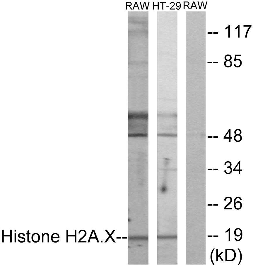
Figure 1. Western blot analysis of HistoneH2A.X using anti-HistoneH2A.X antibody (MP00241). Electrophoresis was performed on a 5-20% SDS-PAGE gel at 70V (Stacking gel) / 90V (Resolving gel) for 2-3 hours. The sample well of each lane was loaded with 30 ug of sample under reducing conditions. Lane 1: human Jurkat whole cell lysates, Lane 2: human U20S whole cell lysates, Lane 3: human PC-3 whole cell lysates, Lane 4: human 293T whole cell lysates, Lane 5: rat PC-12 whole cell lysates, Lane 6: rat C6 whole cell lysates, Lane 7: mouse NIH/3T3 whole cell lysates, Lane 8: mouse Neuro-2a whole cell lysates. After electrophoresis, proteins were transferred to a nitrocellulose membrane at 150 mA for 50-90 minutes. Blocked the membrane with 5% non-fat milk/TBS for 1.5 hour at RT. The membrane was incubated with rabbit anti-HistoneH2A.X antigen affinity purified monoclonal antibody (Catalog # MP00241) at 1:5000 overnight at 4°C, then washed with TBS-0.1%Tween 3 times with 5 minutes each and probed with a goat anti-rabbit IgG-HRP secondary antibody at a dilution of 1:1000 for 1.5 hour at RT. The signal is developed using an Enhanced Chemiluminescent detection (ECL) kit (Catalog # EK1002) with Tanon 5200 system. A specific band was detected for HistoneH2A.X at approximately 15 kDa. The expected band size for HistoneH2A.X is at 15 kDa.
. HistoneH2A.X was detected in a paraffin-embedded section of human colorectal adenocarcinoma tissue. Heat mediated antigen retrieval was performed in EDTA buffer (pH 8.0, epitope retrieval solution). The tissue section was blocked with 10% goat serum. The tissue section was then incubated with 1:50 rabbit anti-HistoneH2A.X Antibody (MP00241) overnight at 4°C. Peroxidase Conjugated Goat Anti-rabbit IgG was used as secondary antibody and incubated for 30 minutes at 37°C. The tissue section was developed using HRP Conjugated Rabbit IgG Super Vision Assay Kit (Catalog # SV0002) with DAB as the chromogen.")
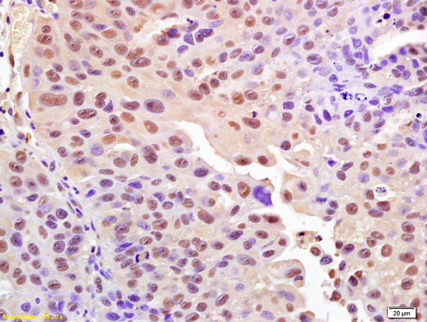
. HistoneH2A.X was detected in a paraffin-embedded section of human liver cancer tissue. Heat mediated antigen retrieval was performed in EDTA buffer (pH 8.0, epitope retrieval solution). The tissue section was blocked with 10% goat serum. The tissue section was then incubated with 1:50 rabbit anti-HistoneH2A.X Antibody (MP00241) overnight at 4°C. Peroxidase Conjugated Goat Anti-rabbit IgG was used as secondary antibody and incubated for 30 minutes at 37°C. The tissue section was developed using HRP Conjugated Rabbit IgG Super Vision Assay Kit (Catalog # SV0002) with DAB as the chromogen.")
. HistoneH2A.X was detected in a paraffin-embedded section of human placenta tissue. Heat mediated antigen retrieval was performed in EDTA buffer (pH 8.0, epitope retrieval solution). The tissue section was blocked with 10% goat serum. The tissue section was then incubated with 1:50 rabbit anti-HistoneH2A.X Antibody (MP00241) overnight at 4°C. Peroxidase Conjugated Goat Anti-rabbit IgG was used as secondary antibody and incubated for 30 minutes at 37°C. The tissue section was developed using HRP Conjugated Rabbit IgG Super Vision Assay Kit (Catalog # SV0002) with DAB as the chromogen.")
. HistoneH2A.X was detected in a paraffin-embedded section of mouse liver tissue. Heat mediated antigen retrieval was performed in EDTA buffer (pH 8.0, epitope retrieval solution). The tissue section was blocked with 10% goat serum. The tissue section was then incubated with 1:50 rabbit anti-HistoneH2A.X Antibody (MP00241) overnight at 4°C. Peroxidase Conjugated Goat Anti-rabbit IgG was used as secondary antibody and incubated for 30 minutes at 37°C. The tissue section was developed using HRP Conjugated Rabbit IgG Super Vision Assay Kit (Catalog # SV0002) with DAB as the chromogen.")
 Antibody.")
Figure 1. Western blot analysis of HistoneH2A.X using anti-HistoneH2A.X antibody (MP00241). Electrophoresis was performed on a 5-20% SDS-PAGE gel at 70V (Stacking gel) / 90V (Resolving gel) for 2-3 hours. The sample well of each lane was loaded with 30 ug of sample under reducing conditions. Lane 1: human Jurkat whole cell lysates, Lane 2: human U20S whole cell lysates, Lane 3: human PC-3 whole cell lysates, Lane 4: human 293T whole cell lysates, Lane 5: rat PC-12 whole cell lysates, Lane 6: rat C6 whole cell lysates, Lane 7: mouse NIH/3T3 whole cell lysates, Lane 8: mouse Neuro-2a whole cell lysates. After electrophoresis, proteins were transferred to a nitrocellulose membrane at 150 mA for 50-90 minutes. Blocked the membrane with 5% non-fat milk/TBS for 1.5 hour at RT. The membrane was incubated with rabbit anti-HistoneH2A.X antigen affinity purified monoclonal antibody (Catalog # MP00241) at 1:5000 overnight at 4°C, then washed with TBS-0.1%Tween 3 times with 5 minutes each and probed with a goat anti-rabbit IgG-HRP secondary antibody at a dilution of 1:1000 for 1.5 hour at RT. The signal is developed using an Enhanced Chemiluminescent detection (ECL) kit (Catalog # EK1002) with Tanon 5200 system. A specific band was detected for HistoneH2A.X at approximately 15 kDa. The expected band size for HistoneH2A.X is at 15 kDa.
Anti-Phospho-Histone H2A.X (S139) H2AFX Monoclonal Antibody
MP00241
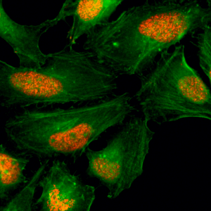
ApplicationsImmunoFluorescence, ImmunoPrecipitation, Western Blot, ImmunoCytoChemistry, ImmunoHistoChemistry
Product group Antibodies
TargetH2AX
Overview
- SupplierBoster Bio
- Product NameAnti-Phospho-Histone H2A.X (S139) Monoclonal Antibody
- Delivery Days Customer9
- ApplicationsImmunoFluorescence, ImmunoPrecipitation, Western Blot, ImmunoCytoChemistry, ImmunoHistoChemistry
- CertificationResearch Use Only
- ClonalityMonoclonal
- Clone IDAbH41
- Gene ID3014
- Target nameH2AX
- Target descriptionH2A.X variant histone
- Target synonymsH2A histone family member X; H2A.X; H2A/X; H2AFX; H2AX histone; histone H2A.x; histone H2AX
- HostRabbit
- IsotypeIgG
- Protein IDP16104
- Protein NameHistone H2AX
- Scientific DescriptionBoster Bio Anti-Phospho-Histone H2A.X (S139) H2AFX Monoclonal Antibody catalog # MP00241. Tested in WB, IHC, ICC/IF, IP applications. This antibody reacts with Human, Mouse, Rat.
- Storage Instruction-20°C
- UNSPSC12352203
References
- Carbamoylation at C-8 position of natural 3-arylcoumarin scaffold for the discovery of novel PARP-1 inhibitors with potent anticancer activity.Read more
- Epoxyeicosatrienoic acids alleviate alveolar epithelial cell senescence by inhibiting mitophagy through NOX4/Nrf2 pathway.Read more
- EETs alleviate alveolar epithelial cell senescence by inhibiting endoplasmic reticulum stress through the Trim25/Keap1/Nrf2 axis.Read more
- Transcriptome Sequencing Reveals Tgf-beta-Mediated Noncoding RNA Regulatory Mechanisms Involved in DNA Damage in the 661W Photoreceptor Cell Line.Read more
- COX-2/sEH Dual Inhibitor Alleviates Hepatocyte Senescence in NAFLD Mice by Restoring Autophagy through Sirt1/PI3K/AKT/mTOR. Zhang CY et al., 2022 Jul 27, Int J Mol SciRead more
Datasheet
MSDS
Related products
Product group Antibodies
Anti-Histone H2AX Antibody144-65468
ApplicationsImmunoFluorescence, Western Blot, ImmunoHistoChemistry
TargetH2AX
- SizePrice
Product group Antibodies
H2Afx Polyclonal AntibodyCAC07288
ApplicationsELISA, ImmunoHistoChemistry
TargetH2AX
- SizePrice
Product group Antibodies

ApplicationsWestern Blot, ELISA, ImmunoHistoChemistry
TargetH2AX
- SizePrice
Product group Antibodies
References
Histone H2A.X antibodyGTX108272
ApplicationsImmunoFluorescence, ImmunoPrecipitation, Western Blot, ImmunoCytoChemistry, ImmunoHistoChemistry, ImmunoHistoChemistry Paraffin
TargetH2AX
- SizePrice
Product group Antibodies

H2AFX / H2AX Antibody (C-Terminus)LS-C358767
ApplicationsImmunoFluorescence, Western Blot, ImmunoCytoChemistry, ImmunoHistoChemistry, ImmunoHistoChemistry Paraffin
TargetH2AX
- SizePrice

Product group Antibodies
Anti-H2AFX AntibodyAMAB91346
ApplicationsImmunoCytoChemistry, ImmunoHistoChemistry
ReactivityHuman
TargetH2AX
- SizePrice
Product group Antibodies
ApplicationsWestern Blot, ELISA, ImmunoHistoChemistry
- SizePrice
Product group Antibodies
References
ApplicationsFlow Cytometry, ImmunoFluorescence, Western Blot, ELISA, ImmunoCytoChemistry, ImmunoHistoChemistry, ImmunoHistoChemistry Frozen, ImmunoHistoChemistry Paraffin
TargetH2AX
- SizePrice
Product group Antibodies
anti-Histone H2AX, Rabbit Monoclonal (RM214)REV-31-1090-00
ApplicationsWestern Blot, ELISA, ImmunoCytoChemistry, Other Application
TargetH2AX
- SizePrice