


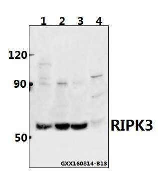
Figure 1. Western blot analysis of Phospho-RIP3 (S232) using anti-Phospho-RIP3 (S232) antibody (M00202S232). Electrophoresis was performed on a 5-20% SDS-PAGE gel at 70V (Stacking gel) / 90V (Resolving gel) for 2-3 hours. The sample well of each lane was loaded with 30 ug of sample under reducing conditions. Lane 1: rat spleen tissue lysates, Lane 2: rat liver tissue lysates, Lane 3: rat heart tissue lysates, Lane 4: rat L6 whole cell lysates, Lane 5: mouse spleen tissue lysates, Lane 6: mouse liver tissue lysates, Lane 7: mouse heart tissue lysates, Lane 8: mouse L929 whole cell lysates. After electrophoresis, proteins were transferred to a nitrocellulose membrane at 150 mA for 50-90 minutes. Blocked the membrane with 5% non-fat milk/TBS for 1.5 hour at RT. The membrane was incubated with rabbit anti-Phospho-RIP3 (S232) antigen affinity purified monoclonal antibody (Catalog # M00202S232) at 1:500 overnight at 4°C, then washed with TBS-0.1%Tween 3 times with 5 minutes each and probed with a goat anti-rabbit IgG-HRP secondary antibody at a dilution of 1:500 for 1.5 hour at RT. The signal is developed using an Enhanced Chemiluminescent detection (ECL) kit (Catalog # EK1002) with Tanon 5200 system. A specific band was detected for Phospho-RIP3 (S232) at approximately 50 kDa. The expected band size for Phospho-RIP3 (S232) is at 50 kDa.
Figure 1. Western blot analysis of Phospho-RIP3 (S232) using anti-Phospho-RIP3 (S232) antibody (M00202S232). Electrophoresis was performed on a 5-20% SDS-PAGE gel at 70V (Stacking gel) / 90V (Resolving gel) for 2-3 hours. The sample well of each lane was loaded with 30 ug of sample under reducing conditions. Lane 1: rat spleen tissue lysates, Lane 2: rat liver tissue lysates, Lane 3: rat heart tissue lysates, Lane 4: rat L6 whole cell lysates, Lane 5: mouse spleen tissue lysates, Lane 6: mouse liver tissue lysates, Lane 7: mouse heart tissue lysates, Lane 8: mouse L929 whole cell lysates. After electrophoresis, proteins were transferred to a nitrocellulose membrane at 150 mA for 50-90 minutes. Blocked the membrane with 5% non-fat milk/TBS for 1.5 hour at RT. The membrane was incubated with rabbit anti-Phospho-RIP3 (S232) antigen affinity purified monoclonal antibody (Catalog # M00202S232) at 1:500 overnight at 4°C, then washed with TBS-0.1%Tween 3 times with 5 minutes each and probed with a goat anti-rabbit IgG-HRP secondary antibody at a dilution of 1:500 for 1.5 hour at RT. The signal is developed using an Enhanced Chemiluminescent detection (ECL) kit (Catalog # EK1002) with Tanon 5200 system. A specific band was detected for Phospho-RIP3 (S232) at approximately 50 kDa. The expected band size for Phospho-RIP3 (S232) is at 50 kDa.
Anti-Phospho-RIP3 (S232) Rabbit Monoclonal Antibody
M00202S232
ApplicationsWestern Blot
Product group Antibodies
ReactivityMouse, Rat
TargetRIPK3
Overview
- SupplierBoster Bio
- Product NameAnti-Phospho-RIP3 (S232) Rabbit Monoclonal Antibody
- Delivery Days Customer9
- ApplicationsWestern Blot
- CertificationResearch Use Only
- ClonalityMonoclonal
- Clone ID31R36
- Gene ID11035
- Target nameRIPK3
- Target descriptionreceptor interacting serine/threonine kinase 3
- Target synonymsRIP3, receptor-interacting serine/threonine-protein kinase 3, RIP-3, RIP-like protein kinase 3, receptor interacting protein 3
- HostRabbit
- IsotypeIgG
- Protein IDQ9Y572
- Protein NameReceptor-interacting serine/threonine-protein kinase 3
- Scientific DescriptionBoster Bio Anti-Phospho-RIP3 (S232) Rabbit Monoclonal Antibody catalog # M00202S232. Tested in WB applications. This antibody reacts with Mouse, Rat.
- ReactivityMouse, Rat
- Storage Instruction-20°C
- UNSPSC12352203
Related products
Product group Antibodies
Anti-RIPK3 AntibodyA28997
ApplicationsWestern Blot
ReactivityHuman, Mouse, Rat
- SizePrice

Product group Antibodies
Anti-RIPK3 AntibodyHPA055087
ApplicationsWestern Blot, ImmunoHistoChemistry
ReactivityHuman
TargetRIPK3
- SizePrice
Product group Antibodies
RIPK3 AntibodyCSB-PA897497ESR1HU
ApplicationsELISA, ImmunoHistoChemistry
ReactivityHuman
TargetRIPK3
- SizePrice
Product group Antibodies

RIPK3 / RIP3 AntibodyLS-C335570
ApplicationsWestern Blot, ImmunoHistoChemistry
ReactivityMouse
TargetRIPK3
- SizePrice
Product group Antibodies
Ripk3 Polyclonal AntibodyCAC09993
ApplicationsImmunoFluorescence, Western Blot, ELISA, ImmunoHistoChemistry
TargetRIPK3
- SizePrice
Product group Antibodies
RIP3 antibodyGTX131188
ApplicationsImmunoFluorescence, Western Blot, ImmunoCytoChemistry, ImmunoHistoChemistry, ImmunoHistoChemistry Paraffin
ReactivityHuman, Rat
TargetRIPK3
- SizePrice
Product group Antibodies
References
RIPK3 Polyclonal AntibodyBS-3551R
ApplicationsImmunoFluorescence, Western Blot, ImmunoHistoChemistry, ImmunoHistoChemistry Frozen, ImmunoHistoChemistry Paraffin
ReactivityHuman, Mouse, Rat
TargetRIPK3
- SizePrice