

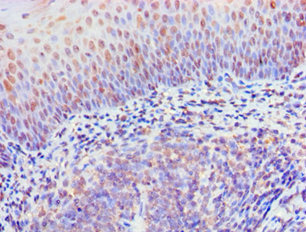
Immunohistochemistry of paraffin-embedded human tonsil tissue using CSB-PA897497ESR1HU at dilution of 1:100


Immunohistochemistry of paraffin-embedded human tonsil tissue using CSB-PA897497ESR1HU at dilution of 1:100
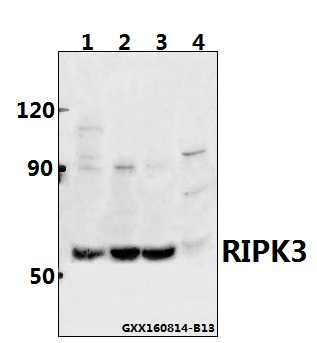
RIPK3 Antibody
CSB-PA897497ESR1HU
ApplicationsELISA, ImmunoHistoChemistry
Product group Antibodies
ReactivityHuman
TargetRIPK3
Overview
- SupplierCusabio
- Product NameRIPK3 Antibody
- Delivery Days Customer20
- ApplicationsELISA, ImmunoHistoChemistry
- CertificationResearch Use Only
- ClonalityPolyclonal
- ConjugateUnconjugated
- Gene ID11035
- Target nameRIPK3
- Target descriptionreceptor interacting serine/threonine kinase 3
- Target synonymsRIP3, receptor-interacting serine/threonine-protein kinase 3, RIP-3, RIP-like protein kinase 3, receptor interacting protein 3
- HostRabbit
- IsotypeIgG
- Protein IDQ9Y572
- Protein NameReceptor-interacting serine/threonine-protein kinase 3
- Scientific DescriptionEssential for necroptosis, a programmed cell death process in response to death-inducing TNF-alpha family members. Upon induction of necrosis, RIPK3 interacts with, and phosphorylates RIPK1 and MLKL to form a necrosis-inducing complex. RIPK3 binds to and enhances the activity of three metabolic enzymes: GLUL, GLUD1, and PYGL. These metabolic enzymes may eventually stimulate the tricarboxylic acid cycle and oxidative phosphorylation, which could result in enhanced ROS production.
- ReactivityHuman
- Storage Instruction-20°C or -80°C
- UNSPSC41116161
Related products
Product group Antibodies
Anti-RIPK3 AntibodyA28997
ApplicationsWestern Blot
ReactivityHuman, Mouse, Rat
- SizePrice

Product group Antibodies
ApplicationsWestern Blot
ReactivityMouse, Rat
TargetRIPK3
- SizePrice

Product group Antibodies
Anti-RIPK3 AntibodyHPA055087
ApplicationsWestern Blot, ImmunoHistoChemistry
ReactivityHuman
TargetRIPK3
- SizePrice
Product group Antibodies

RIPK3 / RIP3 AntibodyLS-C335570
ApplicationsWestern Blot, ImmunoHistoChemistry
ReactivityMouse
TargetRIPK3
- SizePrice
Product group Antibodies
Ripk3 Polyclonal AntibodyCAC09993
ApplicationsImmunoFluorescence, Western Blot, ELISA, ImmunoHistoChemistry
TargetRIPK3
- SizePrice
Product group Antibodies
RIP3 antibodyGTX131188
ApplicationsImmunoFluorescence, Western Blot, ImmunoCytoChemistry, ImmunoHistoChemistry, ImmunoHistoChemistry Paraffin
ReactivityHuman, Rat
TargetRIPK3
- SizePrice
Product group Antibodies
References
RIPK3 Polyclonal AntibodyBS-3551R
ApplicationsImmunoFluorescence, Western Blot, ImmunoHistoChemistry, ImmunoHistoChemistry Frozen, ImmunoHistoChemistry Paraffin
ReactivityHuman, Mouse, Rat
TargetRIPK3
- SizePrice