


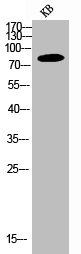
Figure 1. Western blot analysis of PI 3 Kinase p85 alpha/PIK3R1 using anti-PI 3 Kinase p85 alpha/PIK3R1 antibody (A00318-1). Electrophoresis was performed on a 5-20% SDS-PAGE gel at 70V (Stacking gel) / 90V (Resolving gel) for 2-3 hours. The sample well of each lane was loaded with 30 ug of sample under reducing conditions. Lane 1: human Jurkat whole cell lysates, Lane 2: human Hela whole cell lysates, Lane 3: rat PC-12 whole cell lysates, Lane 4: mouse RAW264.7 whole cell lysates. After electrophoresis, proteins were transferred to a nitrocellulose membrane at 150 mA for 50-90 minutes. Blocked the membrane with 5% non-fat milk/TBS for 1.5 hour at RT. The membrane was incubated with rabbit anti-PI 3 Kinase p85 alpha/PIK3R1 antigen affinity purified polyclonal antibody (Catalog # A00318-1) at 0.5 microg/mL overnight at 4°C, then washed with TBS-0.1%Tween 3 times with 5 minutes each and probed with a goat anti-rabbit IgG-HRP secondary antibody at a dilution of 1:5000 for 1.5 hour at RT. The signal is developed using an Enhanced Chemiluminescent detection (ECL) kit (Catalog # EK1002) with Tanon 5200 system. A specific band was detected for PI 3 Kinase p85 alpha/PIK3R1 at approximately 85 kDa. The expected band size for PI 3 Kinase p85 alpha/PIK3R1 is at 85 kDa.
. PI 3 Kinase p85 alpha/PIK3R1 was detected in a paraffin-embedded section of human lung cancer tissue. Heat mediated antigen retrieval was performed in EDTA buffer (pH 8.0, epitope retrieval solution). The tissue section was blocked with 10% goat serum. The tissue section was then incubated with 2 microg/ml rabbit anti-PI 3 Kinase p85 alpha/PIK3R1 Antibody (A00318-1) overnight at 4°C. Biotinylated goat anti-rabbit IgG was used as secondary antibody and incubated for 30 minutes at 37°C. The tissue section was developed using Strepavidin-Biotin-Complex (SABC) (Catalog # SA1022) with DAB as the chromogen.")
. PI 3 Kinase p85 alpha/PIK3R1 was detected in a paraffin-embedded section of human lymphoma tissue. Heat mediated antigen retrieval was performed in EDTA buffer (pH 8.0, epitope retrieval solution). The tissue section was blocked with 10% goat serum. The tissue section was then incubated with 2 microg/ml rabbit anti-PI 3 Kinase p85 alpha/PIK3R1 Antibody (A00318-1) overnight at 4°C. Biotinylated goat anti-rabbit IgG was used as secondary antibody and incubated for 30 minutes at 37°C. The tissue section was developed using Strepavidin-Biotin-Complex (SABC) (Catalog # SA1022) with DAB as the chromogen.")
. PI 3 Kinase p85 alpha/PIK3R1 was detected in a paraffin-embedded section of human testicular cancer tissue. Heat mediated antigen retrieval was performed in EDTA buffer (pH 8.0, epitope retrieval solution). The tissue section was blocked with 10% goat serum. The tissue section was then incubated with 2 microg/ml rabbit anti-PI 3 Kinase p85 alpha/PIK3R1 Antibody (A00318-1) overnight at 4°C. Biotinylated goat anti-rabbit IgG was used as secondary antibody and incubated for 30 minutes at 37°C. The tissue section was developed using Strepavidin-Biotin-Complex (SABC) (Catalog # SA1022) with DAB as the chromogen.")
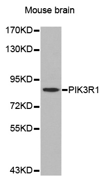
. PI 3 Kinase p85 alpha/PIK3R1 was detected in a paraffin-embedded section of mouse brain tissue. Heat mediated antigen retrieval was performed in EDTA buffer (pH 8.0, epitope retrieval solution). The tissue section was blocked with 10% goat serum. The tissue section was then incubated with 2 microg/ml rabbit anti-PI 3 Kinase p85 alpha/PIK3R1 Antibody (A00318-1) overnight at 4°C. Biotinylated goat anti-rabbit IgG was used as secondary antibody and incubated for 30 minutes at 37°C. The tissue section was developed using Strepavidin-Biotin-Complex (SABC) (Catalog # SA1022) with DAB as the chromogen.")
. PI 3 Kinase p85 alpha/PIK3R1 was detected in a paraffin-embedded section of rat brain tissue. Heat mediated antigen retrieval was performed in EDTA buffer (pH 8.0, epitope retrieval solution). The tissue section was blocked with 10% goat serum. The tissue section was then incubated with 2 microg/ml rabbit anti-PI 3 Kinase p85 alpha/PIK3R1 Antibody (A00318-1) overnight at 4°C. Biotinylated goat anti-rabbit IgG was used as secondary antibody and incubated for 30 minutes at 37°C. The tissue section was developed using Strepavidin-Biotin-Complex (SABC) (Catalog # SA1022) with DAB as the chromogen.")
. PI 3 Kinase p85 alpha/PIK3R1 was detected in an immunocytochemical section of Caco-2 cells. Enzyme antigen retrieval was performed using IHC enzyme antigen retrieval reagent (AR0022) for 15 mins. The cells were blocked with 10% goat serum. And then incubated with 5 microg/mL rabbit anti-PI 3 Kinase p85 alpha/PIK3R1 Antibody (A00318-1) overnight at 4°C. DyLight®488 Conjugated Goat Anti-Rabbit IgG (BA1127) was used as secondary antibody at 1:100 dilution and incubated for 30 minutes at 37°C. The section was counterstained with DAPI. Visualize using a fluorescence microscope and filter sets appropriate for the label used.")
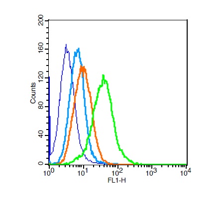
. Overlay histogram showing HL-60 cells stained with A00318-1 (Blue line). To facilitate intracellular staining, cells were fixed with 4% paraformaldehyde and permeabilized with permeabilization buffer. The cells were blocked with 10% normal goat serum. And then incubated with rabbit anti-PI 3 Kinase p85 alpha/PIK3R1 Antibody (A00318-1, 1 microg/1x106 cells) for 30 min at 20°C. DyLight®488 conjugated goat anti-rabbit IgG (BA1127, 5-10 microg/1x106 cells) was used as secondary antibody for 30 minutes at 20°C. Isotype control antibody (Green line) was rabbit IgG (1 microg/1x106) used under the same conditions. Unlabelled sample without incubation with primary antibody and secondary antibody (Red line) was used as a blank control.")
Figure 1. Western blot analysis of PI 3 Kinase p85 alpha/PIK3R1 using anti-PI 3 Kinase p85 alpha/PIK3R1 antibody (A00318-1). Electrophoresis was performed on a 5-20% SDS-PAGE gel at 70V (Stacking gel) / 90V (Resolving gel) for 2-3 hours. The sample well of each lane was loaded with 30 ug of sample under reducing conditions. Lane 1: human Jurkat whole cell lysates, Lane 2: human Hela whole cell lysates, Lane 3: rat PC-12 whole cell lysates, Lane 4: mouse RAW264.7 whole cell lysates. After electrophoresis, proteins were transferred to a nitrocellulose membrane at 150 mA for 50-90 minutes. Blocked the membrane with 5% non-fat milk/TBS for 1.5 hour at RT. The membrane was incubated with rabbit anti-PI 3 Kinase p85 alpha/PIK3R1 antigen affinity purified polyclonal antibody (Catalog # A00318-1) at 0.5 microg/mL overnight at 4°C, then washed with TBS-0.1%Tween 3 times with 5 minutes each and probed with a goat anti-rabbit IgG-HRP secondary antibody at a dilution of 1:5000 for 1.5 hour at RT. The signal is developed using an Enhanced Chemiluminescent detection (ECL) kit (Catalog # EK1002) with Tanon 5200 system. A specific band was detected for PI 3 Kinase p85 alpha/PIK3R1 at approximately 85 kDa. The expected band size for PI 3 Kinase p85 alpha/PIK3R1 is at 85 kDa.
Anti-PI 3 Kinase p85 alpha/PIK3R1 Antibody Picoband(r)
A00318-1-CARRIER-FREE
ApplicationsFlow Cytometry, ImmunoFluorescence, Western Blot, ELISA, ImmunoCytoChemistry, ImmunoHistoChemistry
Product group Antibodies
ReactivityHuman, Mouse, Rat
TargetPIK3R1
Overview
- SupplierBoster Bio
- Product NameAnti-PI 3 Kinase p85 alpha/PIK3R1 Antibody Picoband(r)
- Delivery Days Customer9
- ApplicationsFlow Cytometry, ImmunoFluorescence, Western Blot, ELISA, ImmunoCytoChemistry, ImmunoHistoChemistry
- CertificationResearch Use Only
- ClonalityPolyclonal
- Concentration500 ug/ml
- Gene ID5295
- Target namePIK3R1
- Target descriptionphosphoinositide-3-kinase regulatory subunit 1
- Target synonymsAGM7, GRB1, IMD36, p85, p85-ALPHA, p85alpha, phosphatidylinositol 3-kinase regulatory subunit alpha, PI3 kinase-associated p85, PI3-kinase subunit p85-alpha, PI3K regulatory subunit alpha, growth factor receptor bound 1, phosphatidylinositol 3-kinase 85 kDa regulatory subunit alpha, phosphatidylinositol 3-kinase, regulatory subunit, polypeptide 1 (p85 alpha), phosphatidylinositol 3-kinase-associated p-85 alpha, phosphoinositide-3-kinase regulatory subunit alpha, phosphoinositide-3-kinase, regulatory subunit 1 (alpha), ptdIns-3-kinase regulatory subunit alpha
- HostRabbit
- IsotypeIgG
- Protein IDP27986
- Protein NamePhosphatidylinositol 3-kinase regulatory subunit alpha
- Scientific DescriptionBoster Bio Anti-PI 3 Kinase p85 alpha/PIK3R1 Antibody Picoband® catalog # A00318-1. Tested in ELISA, Flow Cytometry, IF, IHC, ICC, WB applications. This antibody reacts with Human, Mouse, Rat. The brand Picoband indicates this is a premium antibody that guarantees superior quality, high affinity, and strong signals with minimal background in Western blot applications. Only our best-performing antibodies are designated as Picoband, ensuring unmatched performance.
- ReactivityHuman, Mouse, Rat
- Storage Instruction-20°C,2°C to 8°C
- UNSPSC12352203
References
- Volinia S, Patracchini P, Otsu M, et al. Chromosomal localization of human p85 alpha, a subunit of phosphatidylinositol 3-kinase, and its homologue p85 beta. Oncogene. 1992,7(4):789-93.Read this paper
Related products
Product group Antibodies
Anti-PIK3R1 Antibody130-10769
ApplicationsELISA
ReactivityHuman
TargetPIK3R1
- SizePrice
Product group Antibodies
Anti-PIK3R1 AntibodyA42529
ApplicationsWestern Blot
ReactivityHuman, Mouse, Rat
- SizePrice
Product group Antibodies

ApplicationsWestern Blot, ImmunoHistoChemistry, ImmunoHistoChemistry Paraffin
ReactivityMouse, Rat
TargetPIK3R1
- SizePrice
Product group Antibodies
References
PIK3R1 Polyclonal AntibodyBS-0128R
ApplicationsFlow Cytometry, ImmunoFluorescence, Western Blot, ELISA, ImmunoCytoChemistry, ImmunoHistoChemistry, ImmunoHistoChemistry Frozen, ImmunoHistoChemistry Paraffin
ReactivityBovine, Canine, Chicken, Equine, Human, Mouse, Rat
TargetPIK3R1
- SizePrice
Product group Antibodies
PIK3R1 AntibodyCSB-PA003765
ApplicationsWestern Blot, ELISA, ImmunoHistoChemistry
ReactivityHuman, Mouse, Rat
TargetPIK3R1
- SizePrice
Product group Antibodies
Pik3R1 Recombinant AntibodyCAC12508
ApplicationsELISA, ImmunoHistoChemistry
TargetPIK3R1
- SizePrice
Product group Antibodies
Anti-PIK3R1 AntibodyHPA001216
ApplicationsWestern Blot, ImmunoCytoChemistry, ImmunoHistoChemistry
ReactivityHuman
TargetPIK3R1
- SizePrice
![PI3 kinase p85 alpha antibody [N2C1], Internal immunoprecipitates PI3 kinase p85 alpha protein in IP experiments. IP samples: Jurkat whole cell extract A. Control with 5 μg of preimmune Rabbit IgG B. Immunoprecipitation of PI3 kinase p85 alpha protein by 5 μg PI3 kinase p85 alpha antibody [N2C1], Internal (GTX111068) 10 % SDS-PAGE The immunoprecipitated PI3 kinase p85 alpha protein was detected by PI3 kinase p85 alpha antibody [N2C1], Internal (GTX111068) diluted at 1:500. [EasyBlot anti-rabbit IgG (GTX221666-01) was used as a secondary reagent]](https://www.genetex.com/upload/website/prouct_img/normal/GTX111068/GTX111068_40912_IP_w_23060500_860.webp)
Product group Antibodies
ApplicationsImmunoPrecipitation, Western Blot, ImmunoHistoChemistry, ImmunoHistoChemistry Paraffin
ReactivityHuman, Mouse, Rat
TargetPIK3R1
- SizePrice
Product group Antibodies
ApplicationsWestern Blot, ELISA, ImmunoHistoChemistry, ImmunoHistoChemistry Paraffin
ReactivityHuman
TargetPIK3R1
- SizePrice