


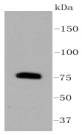
Figure 1. Western blot analysis of PRKCB using anti-PRKCB antibody (A01940). Electrophoresis was performed on a 5-20% SDS-PAGE gel at 70V (Stacking gel) / 90V (Resolving gel) for 2-3 hours. The sample well of each lane was loaded with 30 ug of sample under reducing conditions. Lane 1: human K562 whole cell lysates, Lane 2: human HEL whole cell lysates, Lane 3: human Jurkat whole cell lysates, Lane 4: rat brain tissue lysates, Lane 5: rat PC-12 whole cell lysates, Lane 6: mouse brain tissue lysates. After electrophoresis, proteins were transferred to a nitrocellulose membrane at 150 mA for 50-90 minutes. Blocked the membrane with 5% non-fat milk/TBS for 1.5 hour at RT. The membrane was incubated with rabbit anti-PRKCB antigen affinity purified polyclonal antibody (Catalog # A01940) at 0.5 microg/mL overnight at 4°C, then washed with TBS-0.1%Tween 3 times with 5 minutes each and probed with a goat anti-rabbit IgG-HRP secondary antibody at a dilution of 1:5000 for 1.5 hour at RT. The signal is developed using an Enhanced Chemiluminescent detection (ECL) kit (Catalog # EK1002) with Tanon 5200 system. A specific band was detected for PRKCB at approximately 77 kDa. The expected band size for PRKCB is at 77 kDa.
. PRKCB was detected in a paraffin-embedded section of mouse brain tissue. Heat mediated antigen retrieval was performed in EDTA buffer (pH 8.0, epitope retrieval solution). The tissue section was blocked with 10% goat serum. The tissue section was then incubated with 2 microg/ml rabbit anti-PRKCB Antibody (A01940) overnight at 4°C. Peroxidase Conjugated Goat Anti-rabbit IgG was used as secondary antibody and incubated for 30 minutes at 37°C. The tissue section was developed using HRP Conjugated Rabbit IgG Super Vision Assay Kit (Catalog # SV0002) with DAB as the chromogen.")
. Overlay histogram showing Jurkat cells stained with A01940 (Blue line). To facilitate intracellular staining, cells were fixed with 4% paraformaldehyde and permeabilized with permeabilization buffer. The cells were blocked with 10% normal goat serum. And then incubated with rabbit anti-PRKCB Antibody (A01940, 1 microg/1x106 cells) for 30 min at 20°C. DyLight®488 conjugated goat anti-rabbit IgG (BA1127, 5-10 microg/1x106 cells) was used as secondary antibody for 30 minutes at 20°C. Isotype control antibody (Green line) was rabbit IgG (1 microg/1x106) used under the same conditions. Unlabelled sample without incubation with primary antibody and secondary antibody (Red line) was used as a blank control.")
Figure 1. Western blot analysis of PRKCB using anti-PRKCB antibody (A01940). Electrophoresis was performed on a 5-20% SDS-PAGE gel at 70V (Stacking gel) / 90V (Resolving gel) for 2-3 hours. The sample well of each lane was loaded with 30 ug of sample under reducing conditions. Lane 1: human K562 whole cell lysates, Lane 2: human HEL whole cell lysates, Lane 3: human Jurkat whole cell lysates, Lane 4: rat brain tissue lysates, Lane 5: rat PC-12 whole cell lysates, Lane 6: mouse brain tissue lysates. After electrophoresis, proteins were transferred to a nitrocellulose membrane at 150 mA for 50-90 minutes. Blocked the membrane with 5% non-fat milk/TBS for 1.5 hour at RT. The membrane was incubated with rabbit anti-PRKCB antigen affinity purified polyclonal antibody (Catalog # A01940) at 0.5 microg/mL overnight at 4°C, then washed with TBS-0.1%Tween 3 times with 5 minutes each and probed with a goat anti-rabbit IgG-HRP secondary antibody at a dilution of 1:5000 for 1.5 hour at RT. The signal is developed using an Enhanced Chemiluminescent detection (ECL) kit (Catalog # EK1002) with Tanon 5200 system. A specific band was detected for PRKCB at approximately 77 kDa. The expected band size for PRKCB is at 77 kDa.
Anti-PKC beta 1/PRKCB Antibody Picoband(r)
A01940
ApplicationsFlow Cytometry, Western Blot, ImmunoHistoChemistry
Product group Antibodies
ReactivityHuman, Mouse, Rat
TargetPRKCB
Overview
- SupplierBoster Bio
- Product NameAnti-PKC beta 1/PRKCB Antibody Picoband(r)
- Delivery Days Customer9
- ApplicationsFlow Cytometry, Western Blot, ImmunoHistoChemistry
- CertificationResearch Use Only
- ClonalityPolyclonal
- Concentration500 ug/ml
- Gene ID5579
- Target namePRKCB
- Target descriptionprotein kinase C beta
- Target synonymsPKC-beta, PKCB, PKCI(2), PKCbeta, PRKCB1, PRKCB2, protein kinase C beta type, PKC-B, protein kinase C, beta 1 polypeptide
- HostRabbit
- IsotypeIgG
- Protein IDP05771
- Protein NameProtein kinase C beta type
- Scientific DescriptionBoster Bio Anti-PKC beta 1/PRKCB Antibody Picoband® catalog # A01940. Tested in Flow Cytometry, IHC, WB applications. This antibody reacts with Human, Mouse, Rat. The brand Picoband indicates this is a premium antibody that guarantees superior quality, high affinity, and strong signals with minimal background in Western blot applications. Only our best-performing antibodies are designated as Picoband, ensuring unmatched performance.
- ReactivityHuman, Mouse, Rat
- Storage Instruction-20°C,2°C to 8°C
- UNSPSC12352203
References
- Gan T, Wang Y, Zhao M, et al. 5-(Bis(3-(2-hydroxyethyl)-1H-indol-2-yl)methyl)-2-hydroxybenzoic acid (BHIMHA): showing a strategy of designing drug to block lung metastasis of tumors. Drug Des Devel Ther. 2016,10:711-21. doi: 10.2147/DDDT.S93570Read this paper
- Zhang XM, Chen J, Xia YG, et al. Apoptosis of murine melanoma B16-BL6 cells induced by quercetin targeting mitochondria, inhibiting expression of PKC-alpha and translocating PKC-delta. Cancer Chemother Pharmacol. 2005,55(3):251-62.Read this paper
Datasheet
MSDS
Related products
Product group Antibodies
PRKCB Polyclonal AntibodyCAC15071
ApplicationsImmunoFluorescence, Western Blot, ELISA, ImmunoHistoChemistry
ReactivityRat
TargetPRKCB
- SizePrice
Product group Antibodies
Anti-PKC beta 1/PRKCB Antibody Picoband(r)A01940-CARRIER-FREE
ApplicationsFlow Cytometry, Western Blot, ImmunoHistoChemistry
ReactivityHuman, Mouse, Rat
TargetPRKCB
- SizePrice
Product group Antibodies
Anti-PRKCB Antibody144-62845
ApplicationsImmunoFluorescence, Western Blot, ImmunoHistoChemistry
ReactivityHuman, Mouse, Rat
TargetPRKCB
- SizePrice
Product group Antibodies

PKC beta 2 AntibodyABX025114
ApplicationsWestern Blot, ELISA, ImmunoHistoChemistry
- SizePrice
Product group Antibodies
Anti-PKC beta AntibodyA32391
ApplicationsFlow Cytometry, ImmunoFluorescence, Western Blot, ImmunoCytoChemistry, ImmunoHistoChemistry
ReactivityHuman, Mouse, Rat
- SizePrice
Product group Antibodies

PRKCB / PKC-Beta AntibodyLS-C760925
ApplicationsWestern Blot
ReactivityBovine, Human, Mouse, Rat, Zebra Fish
TargetPRKCB
- SizePrice
Product group Antibodies

Goat anti-PRKCB, BiotinylatedEB11585-B
ApplicationsWestern Blot, ELISA
ReactivityCanine, Human, Rat
TargetPRKCB
- SizePrice
Product group Antibodies
Phospho-PRKCB (S661) AntibodyCSB-PA030241
ApplicationsWestern Blot, ELISA, ImmunoHistoChemistry
ReactivityHuman, Mouse, Rat
TargetPRKCB
- SizePrice