


Figure 1. Western blot analysis of PMCA1 using anti-PMCA1 antibody (M02669). Electrophoresis was performed on a 5-20% SDS-PAGE gel at 70V (Stacking gel) / 90V (Resolving gel) for 2-3 hours. The sample well of each lane was loaded with 30 ug of sample under reducing conditions. Lane 1: human HepG2 whole cell lysates, Lane 2: human U-87MG whole cell lysates, Lane 3: human U251 whole cell lysates, Lane 4: human SH-SY5Y whole cell lysates, Lane 5: rat C6 whole cell lysates, Lane 6: rat RH35 whole cell lysates, Lane 7: mouse Neuro-2a whole cell lysates. After electrophoresis, proteins were transferred to a nitrocellulose membrane at 150 mA for 50-90 minutes. Blocked the membrane with 5% non-fat milk/TBS for 1.5 hour at RT. The membrane was incubated with rabbit anti-PMCA1 antigen affinity purified monoclonal antibody (Catalog # M02669) at 1:500 overnight at 4°C, then washed with TBS-0.1%Tween 3 times with 5 minutes each and probed with a goat anti-rabbit IgG-HRP secondary antibody at a dilution of 1:500 for 1.5 hour at RT. The signal is developed using an Enhanced Chemiluminescent detection (ECL) kit (Catalog # EK1002) with Tanon 5200 system. A specific band was detected for PMCA1 at approximately 250 kDa. The expected band size for PMCA1 is at 135 kDa.
Figure 1. Western blot analysis of PMCA1 using anti-PMCA1 antibody (M02669). Electrophoresis was performed on a 5-20% SDS-PAGE gel at 70V (Stacking gel) / 90V (Resolving gel) for 2-3 hours. The sample well of each lane was loaded with 30 ug of sample under reducing conditions. Lane 1: human HepG2 whole cell lysates, Lane 2: human U-87MG whole cell lysates, Lane 3: human U251 whole cell lysates, Lane 4: human SH-SY5Y whole cell lysates, Lane 5: rat C6 whole cell lysates, Lane 6: rat RH35 whole cell lysates, Lane 7: mouse Neuro-2a whole cell lysates. After electrophoresis, proteins were transferred to a nitrocellulose membrane at 150 mA for 50-90 minutes. Blocked the membrane with 5% non-fat milk/TBS for 1.5 hour at RT. The membrane was incubated with rabbit anti-PMCA1 antigen affinity purified monoclonal antibody (Catalog # M02669) at 1:500 overnight at 4°C, then washed with TBS-0.1%Tween 3 times with 5 minutes each and probed with a goat anti-rabbit IgG-HRP secondary antibody at a dilution of 1:500 for 1.5 hour at RT. The signal is developed using an Enhanced Chemiluminescent detection (ECL) kit (Catalog # EK1002) with Tanon 5200 system. A specific band was detected for PMCA1 at approximately 250 kDa. The expected band size for PMCA1 is at 135 kDa.
Anti-PMCA1 Rabbit Monoclonal Antibody
M02669
ApplicationsWestern Blot, ImmunoHistoChemistry
Product group Antibodies
ReactivityHuman, Mouse, Rat
TargetATP2B1
Overview
- SupplierBoster Bio
- Product NameAnti-PMCA1 Rabbit Monoclonal Antibody
- Delivery Days Customer9
- ApplicationsWestern Blot, ImmunoHistoChemistry
- CertificationResearch Use Only
- ClonalityMonoclonal
- Clone ID22A09
- Gene ID490
- Target nameATP2B1
- Target descriptionATPase plasma membrane Ca2+ transporting 1
- Target synonymsMRD66, PMCA1, PMCA1kb, plasma membrane calcium-transporting ATPase 1, ATPase, Ca++ transporting, plasma membrane 1, plasma membrane calcium pump
- HostRabbit
- IsotypeIgG
- Protein IDP20020
- Protein NamePlasma membrane calcium-transporting ATPase 1
- Scientific DescriptionBoster Bio Anti-PMCA1 Rabbit Monoclonal Antibody catalog # M02669. Tested in WB, IHC applications. This antibody reacts with Human, Mouse, Rat.
- ReactivityHuman, Mouse, Rat
- Storage Instruction-20°C
- UNSPSC12352203
Related products
Product group Antibodies
Anti-ATP2B1 Antibody101-10127
ApplicationsWestern Blot, ELISA
TargetATP2B1
- SizePrice
Product group Antibodies
References
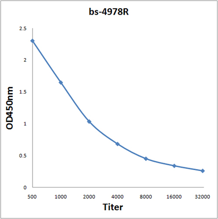
PMCA1 Polyclonal AntibodyBS-4978R
ApplicationsImmunoFluorescence, Western Blot, ELISA, ImmunoCytoChemistry, ImmunoHistoChemistry, ImmunoHistoChemistry Frozen, ImmunoHistoChemistry Paraffin
ReactivityBovine, Canine, Equine, Human, Mouse, Porcine, Rabbit, Rat
TargetATP2B1
- SizePrice
Product group Antibodies

Goat anti-ATP2B1 (aa312-327)EB11182
ApplicationsWestern Blot, ELISA
ReactivityBovine, Canine, Human, Mouse, Porcine, Rat
TargetATP2B1
- SizePrice
Product group Antibodies

ATP2B1 / PMCA1 Antibody (clone 3E2)LS-C196715
ApplicationsWestern Blot, ELISA
ReactivityHuman
TargetATP2B1
- SizePrice

Product group Antibodies
Anti-ATP2B1-25ulHPA011166
ApplicationsWestern Blot, ImmunoCytoChemistry, ImmunoHistoChemistry
ReactivityHuman
- SizePrice
Product group Antibodies
PMCA1 antibodyGTX130858
ApplicationsWestern Blot, ImmunoHistoChemistry, ImmunoHistoChemistry Frozen, ImmunoHistoChemistry Paraffin
ReactivityHuman, Mouse, Rat
TargetATP2B1
- SizePrice