


Immunohistochemical staining of human small intestine shows strong nuclear positivity in glandular cells.

Immunohistochemical staining of human small intestine shows strong nuclear positivity in glandular cells.
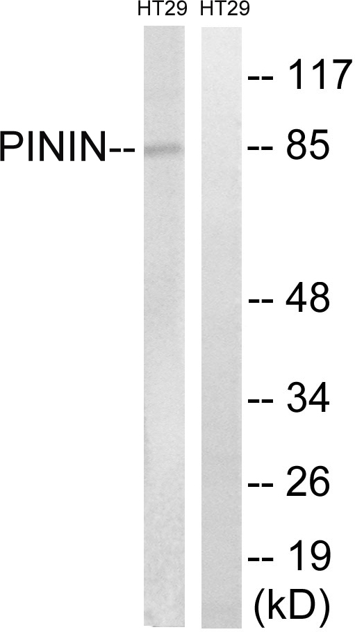
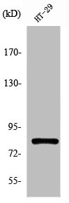
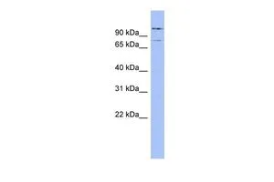
Anti-PNN Antibody
HPA001378
ApplicationsWestern Blot, ImmunoCytoChemistry, ImmunoHistoChemistry
Product group Antibodies
ReactivityHuman
TargetPNN
Overview
- SupplierAtlas Antibodies
- Product NameAnti-PNN Antibody
- Delivery Days Customer4
- ApplicationsWestern Blot, ImmunoCytoChemistry, ImmunoHistoChemistry
- CertificationResearch Use Only
- ClonalityPolyclonal
- ConjugateUnconjugated
- Gene ID5411
- Target namePNN
- Target descriptionpinin, desmosome associated protein
- Target synonymsDRS, DRSP, SDK3, memA, pinin, 140 kDa nuclear and cell adhesion-related phosphoprotein, SR-like protein, desmosome-associated protein, domain-rich serine protein, melanoma metastasis clone A protein, neutrophil protein, nuclear protein SDK3
- HostRabbit
- IsotypeIgG
- Protein IDQ9H307
- Protein NamePinin
- Scientific DescriptionRecombinant Protein Epitope Signature Tag (PrEST) antigen sequence
- ReactivityHuman
- Storage Instruction-20°C,2°C to 8°C
- UNSPSC41116161
Datasheet
MSDS
Related products
Product group Antibodies
Anti-PNN AntibodyA99704
ApplicationsWestern Blot, ELISA
ReactivityHuman, Mouse
- SizePrice

Product group Antibodies
Anti-PNN/DRSP Antibody Picoband(r)A01590-2-CARRIER-FREE
ApplicationsFlow Cytometry, Western Blot, ELISA
ReactivityHuman, Mouse, Rat
TargetPNN
- SizePrice
Product group Antibodies
PNN AntibodyCSB-PA003786
ApplicationsWestern Blot, ELISA
ReactivityHuman, Mouse
TargetPNN
- SizePrice
Product group Antibodies

PNN / Pinin AntibodyLS-C403434
ApplicationsELISA, ImmunoHistoChemistry
ReactivityHuman, Mouse
TargetPNN
- SizePrice
Product group Antibodies
PNN antibody, InternalGTX45055
ApplicationsWestern Blot
ReactivityHuman
TargetPNN
- SizePrice