


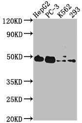
Figure 1. Western blot analysis of POGLUT1 using anti-POGLUT1 antibody (A06826-1). Electrophoresis was performed on a 5-20% SDS-PAGE gel at 70V (Stacking gel) / 90V (Resolving gel) for 2-3 hours. The sample well of each lane was loaded with 30 ug of sample under reducing conditions. Lane 1: human HL-60 whole cell lysates, Lane 2: human HepG2 whole cell lysates, Lane 3: human U87 whole cell lysates, Lane 4: human Raji whole cell lysates, Lane 5: human Jurkat whole cell lysates, Lane 6: human Hela whole cell lysates, Lane 7: mouse thymus tissue lysates, Lane 8: mouse Ana-1 whole cell lysates. After electrophoresis, proteins were transferred to a nitrocellulose membrane at 150 mA for 50-90 minutes. Blocked the membrane with 5% non-fat milk/TBS for 1.5 hour at RT. The membrane was incubated with rabbit anti-POGLUT1 antigen affinity purified polyclonal antibody (Catalog # A06826-1) at 0.5 microg/mL overnight at 4°C, then washed with TBS-0.1%Tween 3 times with 5 minutes each and probed with a goat anti-rabbit IgG-HRP secondary antibody at a dilution of 1:5000 for 1.5 hour at RT. The signal is developed using an Enhanced Chemiluminescent detection (ECL) kit (Catalog # EK1002) with Tanon 5200 system. A specific band was detected for POGLUT1 at approximately 55 kDa. The expected band size for POGLUT1 is at 55 kDa.
. POGLUT1 was detected in a paraffin-embedded section of human breast cancer tissue. Heat mediated antigen retrieval was performed in EDTA buffer (pH 8.0, epitope retrieval solution). The tissue section was blocked with 10% goat serum. The tissue section was then incubated with 2 microg/ml rabbit anti-POGLUT1 Antibody (A06826-1) overnight at 4°C. Biotinylated goat anti-rabbit IgG was used as secondary antibody and incubated for 30 minutes at 37°C. The tissue section was developed using Strepavidin-Biotin-Complex (SABC) (Catalog # SA1022) with DAB as the chromogen.")
. POGLUT1 was detected in a paraffin-embedded section of human gallbladder adenocarcinoma tissue. Heat mediated antigen retrieval was performed in EDTA buffer (pH 8.0, epitope retrieval solution). The tissue section was blocked with 10% goat serum. The tissue section was then incubated with 2 microg/ml rabbit anti-POGLUT1 Antibody (A06826-1) overnight at 4°C. Biotinylated goat anti-rabbit IgG was used as secondary antibody and incubated for 30 minutes at 37°C. The tissue section was developed using Strepavidin-Biotin-Complex (SABC) (Catalog # SA1022) with DAB as the chromogen.")
. POGLUT1 was detected in a paraffin-embedded section of human gastric cancer tissue. Heat mediated antigen retrieval was performed in EDTA buffer (pH 8.0, epitope retrieval solution). The tissue section was blocked with 10% goat serum. The tissue section was then incubated with 2 microg/ml rabbit anti-POGLUT1 Antibody (A06826-1) overnight at 4°C. Biotinylated goat anti-rabbit IgG was used as secondary antibody and incubated for 30 minutes at 37°C. The tissue section was developed using Strepavidin-Biotin-Complex (SABC) (Catalog # SA1022) with DAB as the chromogen.")
. POGLUT1 was detected in a paraffin-embedded section of human lung cancer tissue. Heat mediated antigen retrieval was performed in EDTA buffer (pH 8.0, epitope retrieval solution). The tissue section was blocked with 10% goat serum. The tissue section was then incubated with 2 microg/ml rabbit anti-POGLUT1 Antibody (A06826-1) overnight at 4°C. Biotinylated goat anti-rabbit IgG was used as secondary antibody and incubated for 30 minutes at 37°C. The tissue section was developed using Strepavidin-Biotin-Complex (SABC) (Catalog # SA1022) with DAB as the chromogen.")
. POGLUT1 was detected in a paraffin-embedded section of human lymphoma tissue. Heat mediated antigen retrieval was performed in EDTA buffer (pH 8.0, epitope retrieval solution). The tissue section was blocked with 10% goat serum. The tissue section was then incubated with 2 microg/ml rabbit anti-POGLUT1 Antibody (A06826-1) overnight at 4°C. Biotinylated goat anti-rabbit IgG was used as secondary antibody and incubated for 30 minutes at 37°C. The tissue section was developed using Strepavidin-Biotin-Complex (SABC) (Catalog # SA1022) with DAB as the chromogen.")
. POGLUT1 was detected in a paraffin-embedded section of human ovarian cancer tissue. Heat mediated antigen retrieval was performed in EDTA buffer (pH 8.0, epitope retrieval solution). The tissue section was blocked with 10% goat serum. The tissue section was then incubated with 2 microg/ml rabbit anti-POGLUT1 Antibody (A06826-1) overnight at 4°C. Biotinylated goat anti-rabbit IgG was used as secondary antibody and incubated for 30 minutes at 37°C. The tissue section was developed using Strepavidin-Biotin-Complex (SABC) (Catalog # SA1022) with DAB as the chromogen.")
. POGLUT1 was detected in a paraffin-embedded section of human placenta tissue. Heat mediated antigen retrieval was performed in EDTA buffer (pH 8.0, epitope retrieval solution). The tissue section was blocked with 10% goat serum. The tissue section was then incubated with 2 microg/ml rabbit anti-POGLUT1 Antibody (A06826-1) overnight at 4°C. Biotinylated goat anti-rabbit IgG was used as secondary antibody and incubated for 30 minutes at 37°C. The tissue section was developed using Strepavidin-Biotin-Complex (SABC) (Catalog # SA1022) with DAB as the chromogen.")
. POGLUT1 was detected in an immunocytochemical section of T-47D cells. Enzyme antigen retrieval was performed using IHC enzyme antigen retrieval reagent (AR0022) for 15 mins. The cells were blocked with 10% goat serum. And then incubated with 5 microg/mL rabbit anti-POGLUT1 Antibody (A06826-1) overnight at 4°C. DyLight®488 Conjugated Goat Anti-Rabbit IgG (BA1127) was used as secondary antibody at 1:100 dilution and incubated for 30 minutes at 37°C. The section was counterstained with DAPI. Visualize using a fluorescence microscope and filter sets appropriate for the label used.")
Figure 1. Western blot analysis of POGLUT1 using anti-POGLUT1 antibody (A06826-1). Electrophoresis was performed on a 5-20% SDS-PAGE gel at 70V (Stacking gel) / 90V (Resolving gel) for 2-3 hours. The sample well of each lane was loaded with 30 ug of sample under reducing conditions. Lane 1: human HL-60 whole cell lysates, Lane 2: human HepG2 whole cell lysates, Lane 3: human U87 whole cell lysates, Lane 4: human Raji whole cell lysates, Lane 5: human Jurkat whole cell lysates, Lane 6: human Hela whole cell lysates, Lane 7: mouse thymus tissue lysates, Lane 8: mouse Ana-1 whole cell lysates. After electrophoresis, proteins were transferred to a nitrocellulose membrane at 150 mA for 50-90 minutes. Blocked the membrane with 5% non-fat milk/TBS for 1.5 hour at RT. The membrane was incubated with rabbit anti-POGLUT1 antigen affinity purified polyclonal antibody (Catalog # A06826-1) at 0.5 microg/mL overnight at 4°C, then washed with TBS-0.1%Tween 3 times with 5 minutes each and probed with a goat anti-rabbit IgG-HRP secondary antibody at a dilution of 1:5000 for 1.5 hour at RT. The signal is developed using an Enhanced Chemiluminescent detection (ECL) kit (Catalog # EK1002) with Tanon 5200 system. A specific band was detected for POGLUT1 at approximately 55 kDa. The expected band size for POGLUT1 is at 55 kDa.
Anti-POGLUT1 Antibody Picoband(r)
A06826-1-CARRIER-FREE
ApplicationsImmunoFluorescence, Western Blot, ImmunoCytoChemistry, ImmunoHistoChemistry
Product group Antibodies
ReactivityHuman, Mouse
TargetPOGLUT1
Overview
- SupplierBoster Bio
- Product NameAnti-POGLUT1 Antibody Picoband(r)
- Delivery Days Customer9
- ApplicationsImmunoFluorescence, Western Blot, ImmunoCytoChemistry, ImmunoHistoChemistry
- CertificationResearch Use Only
- ClonalityPolyclonal
- Concentration500 ug/ml
- Gene ID56983
- Target namePOGLUT1
- Target descriptionprotein O-glucosyltransferase 1
- Target synonymsC3orf9, CLP46, KDELCL1, KTELC1, LGMD2Z, LGMDR21, MDS010, MDSRP, Rumi, hCLP46, protein O-glucosyltransferase 1, 9630046K23Rik, CAP10-like 46 kDa protein, KDELC family like 1, KTEL (Lys-Tyr-Glu-Leu) containing 1, KTEL motif-containing protein 1, O-glucosyltransferase Rumi homolog, hRumi, myelodysplastic syndromes relative protein, protein O-xylosyltransferase POGLUT1, x 010 protein
- HostRabbit
- IsotypeIgG
- Protein IDQ8NBL1
- Protein NameProtein O-glucosyltransferase 1
- Scientific DescriptionBoster Bio Anti-POGLUT1 Antibody Picoband® catalog # A06826-1. Tested in IF, IHC, ICC, WB applications. This antibody reacts with Human, Mouse. The brand Picoband indicates this is a premium antibody that guarantees superior quality, high affinity, and strong signals with minimal background in Western blot applications. Only our best-performing antibodies are designated as Picoband, ensuring unmatched performance.
- ReactivityHuman, Mouse
- Storage Instruction-20°C,2°C to 8°C
- UNSPSC12352203
Related products
Product group Antibodies
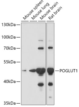
Anti-POGLUT1 AntibodyA308011
ApplicationsImmunoFluorescence, Western Blot, ImmunoCytoChemistry
ReactivityMouse, Rat
- SizePrice
Product group Antibodies

POGLUT1 AntibodyLS-C755103
ApplicationsImmunoFluorescence, Western Blot, ELISA, ImmunoHistoChemistry
ReactivityHuman
TargetPOGLUT1
- SizePrice

Product group Antibodies
Anti-POGLUT1 AntibodyHPA037855
ApplicationsWestern Blot, ImmunoHistoChemistry
ReactivityHuman
TargetPOGLUT1
- SizePrice
Product group Antibodies
POGLUT1 AntibodyCSB-PA818754LA01HU
ApplicationsWestern Blot, ELISA, ImmunoHistoChemistry
ReactivityHuman
TargetPOGLUT1
- SizePrice
Product group Antibodies
POGLUT1 Polyclonal AntibodyCAC15850
ApplicationsWestern Blot, ELISA, ImmunoHistoChemistry
TargetPOGLUT1
- SizePrice
Product group Antibodies
Anti-POGLUT1 Antibody144-65126
ApplicationsImmunoFluorescence, Western Blot
ReactivityHuman, Mouse, Rat
TargetPOGLUT1
- SizePrice
Product group Antibodies
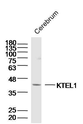
KTEL1 Polyclonal AntibodyBS-16861R
ApplicationsImmunoFluorescence, Western Blot, ELISA, ImmunoCytoChemistry, ImmunoHistoChemistry, ImmunoHistoChemistry Frozen, ImmunoHistoChemistry Paraffin
ReactivityBovine, Canine, Equine, Human, Mouse, Rabbit, Rat, Sheep
TargetPOGLUT1
- SizePrice