


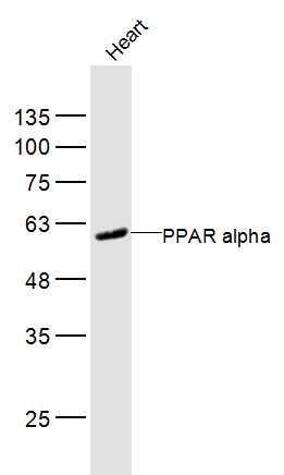
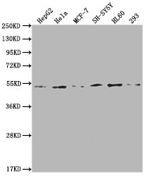
Figure 1. Western blot analysis of PPAR alpha/PPARA using anti-PPAR alpha/PPARA antibody (A00600-2). Electrophoresis was performed on a 5-20% SDS-PAGE gel at 70V (Stacking gel) / 90V (Resolving gel) for 2-3 hours. The sample well of each lane was loaded with 30 ug of sample under reducing conditions. Lane 1: human HepG2 whole cell lysates, Lane 2: human Hela whole cell lysates, Lane 3: human MCF-7 whole cell lysates, Lane 4: rat liver tissue lysates, Lane 5: mouse liver tissue lysates. After electrophoresis, proteins were transferred to a nitrocellulose membrane at 150 mA for 50-90 minutes. Blocked the membrane with 5% non-fat milk/TBS for 1.5 hour at RT. The membrane was incubated with rabbit anti-PPAR alpha/PPARA antigen affinity purified polyclonal antibody (Catalog # A00600-2) at 0.5 microg/mL overnight at 4°C, then washed with TBS-0.1%Tween 3 times with 5 minutes each and probed with a goat anti-rabbit IgG-HRP secondary antibody at a dilution of 1:5000 for 1.5 hour at RT. The signal is developed using an Enhanced Chemiluminescent detection (ECL) kit (Catalog # EK1002) with Tanon 5200 system. A specific band was detected for PPAR alpha/PPARA at approximately 52 kDa. The expected band size for PPAR alpha/PPARA is at 52 kDa.
. Overlay histogram showing HepG2 cells stained with A00600-2 (Blue line). To facilitate intracellular staining, cells were fixed with 4% paraformaldehyde and permeabilized with permeabilization buffer. The cells were blocked with 10% normal goat serum. And then incubated with rabbit anti-PPAR alpha/PPARA Antibody (A00600-2, 1 microg/1x106 cells) for 30 min at 20°C. DyLight®488 conjugated goat anti-rabbit IgG (BA1127, 5-10 microg/1x106 cells) was used as secondary antibody for 30 minutes at 20°C. Isotype control antibody (Green line) was rabbit IgG (1 microg/1x106) used under the same conditions. Unlabelled sample without incubation with primary antibody and secondary antibody (Red line) was used as a blank control.")
Figure 1. Western blot analysis of PPAR alpha/PPARA using anti-PPAR alpha/PPARA antibody (A00600-2). Electrophoresis was performed on a 5-20% SDS-PAGE gel at 70V (Stacking gel) / 90V (Resolving gel) for 2-3 hours. The sample well of each lane was loaded with 30 ug of sample under reducing conditions. Lane 1: human HepG2 whole cell lysates, Lane 2: human Hela whole cell lysates, Lane 3: human MCF-7 whole cell lysates, Lane 4: rat liver tissue lysates, Lane 5: mouse liver tissue lysates. After electrophoresis, proteins were transferred to a nitrocellulose membrane at 150 mA for 50-90 minutes. Blocked the membrane with 5% non-fat milk/TBS for 1.5 hour at RT. The membrane was incubated with rabbit anti-PPAR alpha/PPARA antigen affinity purified polyclonal antibody (Catalog # A00600-2) at 0.5 microg/mL overnight at 4°C, then washed with TBS-0.1%Tween 3 times with 5 minutes each and probed with a goat anti-rabbit IgG-HRP secondary antibody at a dilution of 1:5000 for 1.5 hour at RT. The signal is developed using an Enhanced Chemiluminescent detection (ECL) kit (Catalog # EK1002) with Tanon 5200 system. A specific band was detected for PPAR alpha/PPARA at approximately 52 kDa. The expected band size for PPAR alpha/PPARA is at 52 kDa.
Anti-PPAR alpha/PPARA Antibody Picoband(r)
A00600-2-CARRIER-FREE
ApplicationsFlow Cytometry, Western Blot, ELISA
Product group Antibodies
ReactivityHuman, Mouse, Rat
TargetPPARA
Overview
- SupplierBoster Bio
- Product NameAnti-PPAR alpha/PPARA Antibody Picoband(r)
- Delivery Days Customer9
- ApplicationsFlow Cytometry, Western Blot, ELISA
- CertificationResearch Use Only
- ClonalityPolyclonal
- Concentration500 ug/ml
- Gene ID5465
- Target namePPARA
- Target descriptionperoxisome proliferator activated receptor alpha
- Target synonymsNR1C1, PPAR, PPAR-alpha, PPARalpha, hPPAR, peroxisome proliferator-activated receptor alpha, nuclear receptor subfamily 1 group C member 1, peroxisome proliferative activated receptor, alpha
- HostRabbit
- IsotypeIgG
- Protein IDQ07869
- Protein NamePeroxisome proliferator-activated receptor alpha
- Scientific DescriptionBoster Bio Anti-PPAR alpha/PPARA Antibody Picoband® catalog # A00600-2. Tested in ELISA, Flow Cytometry, WB applications. This antibody reacts with Human, Mouse, Rat. The brand Picoband indicates this is a premium antibody that guarantees superior quality, high affinity, and strong signals with minimal background in Western blot applications. Only our best-performing antibodies are designated as Picoband, ensuring unmatched performance.
- ReactivityHuman, Mouse, Rat
- Storage Instruction-20°C,2°C to 8°C
- UNSPSC12352203
References
- Kersten S, Seydoux J, Peters JM, et al. Peroxisome proliferator-activated receptor alpha mediates the adaptive response to fasting. J Clin Invest. 1999,103(11):1489-98.Read this paper
- Sher T, Yi HF, McBride OW, et al. cDNA cloning, chromosomal mapping, and functional characterization of the human peroxisome proliferator activated receptor. Biochemistry. 1993,32(21):5598-604.Read this paper
Related products
Product group Antibodies
Anti-PPAR alpha AntibodyA283081
ApplicationsWestern Blot, ELISA
ReactivityHuman
- SizePrice
Product group Antibodies
References
PPAR alpha Polyclonal AntibodyBS-23398R
ApplicationsImmunoFluorescence, Western Blot, ELISA, ImmunoCytoChemistry, ImmunoHistoChemistry, ImmunoHistoChemistry Frozen, ImmunoHistoChemistry Paraffin
ReactivityCanine, Chicken, Equine, Human, Mouse, Porcine, Rabbit, Rat, Sheep
TargetPPARA
- SizePrice
Product group Antibodies
PPARA Polyclonal AntibodyCAC15863
ApplicationsWestern Blot, ELISA
TargetPPARA
- SizePrice
Product group Antibodies
PPARA AntibodyCSB-PA018421LA01HU
ApplicationsWestern Blot, ELISA
ReactivityHuman
TargetPPARA
- SizePrice
Product group Antibodies
PPAR alpha antibodyGTX101098
ApplicationsImmunoPrecipitation, Western Blot, ChIP Chromatin ImmunoPrecipitation, ImmunoHistoChemistry, ImmunoHistoChemistry Paraffin
ReactivityHuman, Insect, Mouse, Plant, Rat
TargetPPARA
- SizePrice

Product group Antibodies
Anti-PPARA AntibodyHPA058901
ApplicationsImmunoCytoChemistry
ReactivityHuman
TargetPPARA
- SizePrice
Product group Antibodies

PPARA / PPAR Alpha AntibodyLS-C769771
ApplicationsWestern Blot
ReactivityHuman
TargetPPARA
- SizePrice