


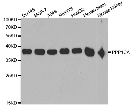
Figure 1. Western blot analysis of PPP1CA + PPP1CB using anti-PPP1CA + PPP1CB antibody (M02801). Electrophoresis was performed on a 5-20% SDS-PAGE gel at 70V (Stacking gel) / 90V (Resolving gel) for 2-3 hours. The sample well of each lane was loaded with 30 ug of sample under reducing conditions. Lane 1: human Hela whole cell lysates, Lane 2: human Jurkat whole cell lysates, Lane 3: human K562 whole cell lysates, Lane 4: human U87 whole cell lysates, Lane 5: rat brain tissue lysates, Lane 6: rat C6 whole cell lysates, Lane 7: mouse brain tissue lysates, Lane 8: mouse NIH/3T3 whole cell lysates. After electrophoresis, proteins were transferred to a nitrocellulose membrane at 150 mA for 50-90 minutes. Blocked the membrane with 5% non-fat milk/TBS for 1.5 hour at RT. The membrane was incubated with rabbit anti-PPP1CA + PPP1CB antigen affinity purified monoclonal antibody (Catalog # M02801) at 1:500 overnight at 4°C, then washed with TBS-0.1%Tween 3 times with 5 minutes each and probed with a goat anti-rabbit IgG-HRP secondary antibody at a dilution of 1:5000 for 1.5 hour at RT. The signal is developed using an Enhanced Chemiluminescent detection (ECL) kit (Catalog # EK1002) with Tanon 5200 system. A specific band was detected for PPP1CA + PPP1CB at approximately 38 kDa. The expected band size for PPP1CA + PPP1CB is at 38 kDa.
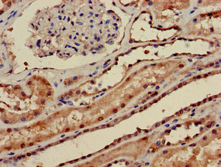
 PPP1CA was detected in paraffin-embedded tissue section. Heat mediated antigen retrieval was performed in citrate buffer (pH6, epitope retrieval solution) for 20 mins. The tissue section was blocked with 10% goat serum. The tissue section was then incubated with 1ug/ml rabbit anti-PPP1CA Antibody (M02801)overnight at 4 Biotinylated goat anti-rabbit IgG was used as secondary antibody and incubated for 30 minutes at 37 The tissue section was developed using Strepavidin-Biotin-Complex (SABC)(Catalog # SA1022) with DAB as the chromogen.")
Figure 1. Western blot analysis of PPP1CA + PPP1CB using anti-PPP1CA + PPP1CB antibody (M02801). Electrophoresis was performed on a 5-20% SDS-PAGE gel at 70V (Stacking gel) / 90V (Resolving gel) for 2-3 hours. The sample well of each lane was loaded with 30 ug of sample under reducing conditions. Lane 1: human Hela whole cell lysates, Lane 2: human Jurkat whole cell lysates, Lane 3: human K562 whole cell lysates, Lane 4: human U87 whole cell lysates, Lane 5: rat brain tissue lysates, Lane 6: rat C6 whole cell lysates, Lane 7: mouse brain tissue lysates, Lane 8: mouse NIH/3T3 whole cell lysates. After electrophoresis, proteins were transferred to a nitrocellulose membrane at 150 mA for 50-90 minutes. Blocked the membrane with 5% non-fat milk/TBS for 1.5 hour at RT. The membrane was incubated with rabbit anti-PPP1CA + PPP1CB antigen affinity purified monoclonal antibody (Catalog # M02801) at 1:500 overnight at 4°C, then washed with TBS-0.1%Tween 3 times with 5 minutes each and probed with a goat anti-rabbit IgG-HRP secondary antibody at a dilution of 1:5000 for 1.5 hour at RT. The signal is developed using an Enhanced Chemiluminescent detection (ECL) kit (Catalog # EK1002) with Tanon 5200 system. A specific band was detected for PPP1CA + PPP1CB at approximately 38 kDa. The expected band size for PPP1CA + PPP1CB is at 38 kDa.
Anti-PPP1CA+PPP1CB Rabbit Monoclonal Antibody
M02801
ApplicationsImmunoFluorescence, ImmunoPrecipitation, Western Blot, ImmunoCytoChemistry, ImmunoHistoChemistry
Product group Antibodies
ReactivityHuman, Mouse, Rat
TargetPPP1CA
Overview
- SupplierBoster Bio
- Product NameAnti-PPP1CA+PPP1CB Rabbit Monoclonal Antibody
- Delivery Days Customer9
- ApplicationsImmunoFluorescence, ImmunoPrecipitation, Western Blot, ImmunoCytoChemistry, ImmunoHistoChemistry
- CertificationResearch Use Only
- ClonalityMonoclonal
- Clone IDAFC-16
- Gene ID5499
- Target namePPP1CA
- Target descriptionprotein phosphatase 1 catalytic subunit alpha
- Target synonymsPP-1A, PP1A, PP1alpha, PPP1A, serine/threonine-protein phosphatase PP1-alpha catalytic subunit, protein phosphatase 1, catalytic subunit, alpha isozyme, serine/threonine protein phosphatase PP1-alpha 1 catalytic subunit, testicular tissue protein Li 155
- HostRabbit
- IsotypeIgG
- Protein IDP62136
- Protein NameSerine/threonine-protein phosphatase PP1-alpha catalytic subunit
- Scientific DescriptionBoster Bio Anti-PPP1CA+PPP1CB Rabbit Monoclonal Antibody catalog # M02801. Tested in WB, IHC, ICC/IF, IP applications. This antibody reacts with Human, Mouse, Rat.
- ReactivityHuman, Mouse, Rat
- Storage Instruction-20°C
- UNSPSC12352203
References
- Liu TC, Li HX, Wan YX, et al. METTL14-mediated upregulation of lncRNA HOTAIR represses PP1α expression by promoting H3K4me1 demethylation in oxycodone-treated mice. CNS Neurosci Ther. 2024,30(7):e14830. doi: 10.1111/cns.14830Read this paper
Datasheet
MSDS
Related products
Product group Antibodies
Anti-PPP1CA AntibodyA29955
ApplicationsImmunoFluorescence, Western Blot, ImmunoHistoChemistry
ReactivityHuman, Mouse, Rat
- SizePrice

Product group Antibodies
Anti-PPP1CA Antibody144-12468
ApplicationsWestern Blot, ImmunoHistoChemistry
ReactivityHuman, Mouse
TargetPPP1CA
- SizePrice
Product group Antibodies

PPP1CA / PP1-Alpha AntibodyLS-C747567
ApplicationsWestern Blot, ImmunoHistoChemistry
ReactivityHuman, Mouse
TargetPPP1CA
- SizePrice
Product group Antibodies
PPP1CA Polyclonal AntibodyBS-3756R
ApplicationsImmunoFluorescence, ELISA, ImmunoCytoChemistry, ImmunoHistoChemistry, ImmunoHistoChemistry Frozen, ImmunoHistoChemistry Paraffin
ReactivityCanine, Equine, Human, Mouse, Rabbit, Rat
TargetPPP1CA
- SizePrice
Product group Antibodies
ApplicationsImmunoPrecipitation, Western Blot, ImmunoCytoChemistry, ImmunoHistoChemistry
ReactivityMouse, Porcine, Rat
TargetPPP1CA
- SizePrice
Product group Antibodies
PPP1CA AntibodyCSB-PA018503LA01HU
ApplicationsImmunoFluorescence, ELISA, ImmunoHistoChemistry
ReactivityHuman
TargetPPP1CA
- SizePrice

Product group Antibodies
Anti-PPP1CA AntibodyHPA046833
ApplicationsWestern Blot, ImmunoCytoChemistry, ImmunoHistoChemistry
ReactivityHuman
TargetPPP1CA
- SizePrice
Product group Antibodies
PPP1A antibodyGTX105255
ApplicationsImmunoFluorescence, ImmunoPrecipitation, Western Blot, ImmunoCytoChemistry, ImmunoHistoChemistry, ImmunoHistoChemistry Paraffin
ReactivityHuman, Mouse, Rat, Zebra Fish
TargetPPP1CA
- SizePrice