

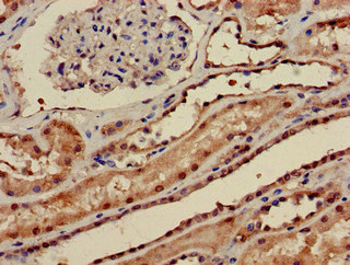
Immunohistochemistry of paraffin-embedded human kidney tissue using CSB-PA018503LA01HU at dilution of 1:100

")
Immunohistochemistry of paraffin-embedded human kidney tissue using CSB-PA018503LA01HU at dilution of 1:100
PPP1CA Antibody
CSB-PA018503LA01HU
ApplicationsImmunoFluorescence, ELISA, ImmunoHistoChemistry
Product group Antibodies
ReactivityHuman
TargetPPP1CA
Overview
- SupplierCusabio
- Product NamePPP1CA Antibody
- Delivery Days Customer20
- ApplicationsImmunoFluorescence, ELISA, ImmunoHistoChemistry
- CertificationResearch Use Only
- ClonalityPolyclonal
- ConjugateUnconjugated
- Gene ID5499
- Target namePPP1CA
- Target descriptionprotein phosphatase 1 catalytic subunit alpha
- Target synonymsPP-1A, PP1A, PP1alpha, PPP1A, serine/threonine-protein phosphatase PP1-alpha catalytic subunit, protein phosphatase 1, catalytic subunit, alpha isozyme, serine/threonine protein phosphatase PP1-alpha 1 catalytic subunit, testicular tissue protein Li 155
- HostRabbit
- IsotypeIgG
- Protein IDP62136
- Protein NameSerine/threonine-protein phosphatase PP1-alpha catalytic subunit
- Scientific DescriptionProtein phosphatase that associates with over 200 regulatory proteins to form highly specific holoenzymes which dephosphorylate hundreds of biological targets. Protein phosphatase 1 (PP1) is essential for cell division, and participates in the regulation of glycogen metabolism, muscle contractility and protein synthesis. Involved in regulation of ionic conductances and long-term synaptic plasticity. May play an important role in dephosphorylating substrates such as the postsynaptic density-associated Ca(2+)/calmodulin dependent protein kinase II. Component of the PTW/PP1 phosphatase complex, which plays a role in the control of chromatin structure and cell cycle progression during the transition from mitosis into interphase. Regulates NEK2 function in terms of kinase activity and centrosome number and splitting, both in the presence and absence of radiation-induced DNA damage. Regulator of neural tube and optic fissure closure, and enteric neural crest cell (ENCCs) migration during development. In balance with CSNK1D and CSNK1E, determines the circadian period length, through the regulation of the speed and rhythmicity of PER1 and PER2 phosphorylation. May dephosphorylate CSNK1D and CSNK1E. Dephosphorylates the Ser-418 residue of FOXP3 in regulatory T-cells (Treg) from patients with rheumatoid arthritis, thereby inactivating FOXP3 and rendering Treg cells functionally defective (PubMed:23396208).
- ReactivityHuman
- Storage Instruction-20°C or -80°C
- UNSPSC41116161
Related products
Product group Antibodies
Anti-PPP1CA AntibodyA29955
ApplicationsImmunoFluorescence, Western Blot, ImmunoHistoChemistry
ReactivityHuman, Mouse, Rat
- SizePrice

Product group Antibodies
Anti-PPP1CA Antibody144-12468
ApplicationsWestern Blot, ImmunoHistoChemistry
ReactivityHuman, Mouse
TargetPPP1CA
- SizePrice
Product group Antibodies

PPP1CA / PP1-Alpha AntibodyLS-C747567
ApplicationsWestern Blot, ImmunoHistoChemistry
ReactivityHuman, Mouse
TargetPPP1CA
- SizePrice
Product group Antibodies
PPP1CA Polyclonal AntibodyBS-3756R
ApplicationsImmunoFluorescence, ELISA, ImmunoCytoChemistry, ImmunoHistoChemistry, ImmunoHistoChemistry Frozen, ImmunoHistoChemistry Paraffin
ReactivityCanine, Equine, Human, Mouse, Rabbit, Rat
TargetPPP1CA
- SizePrice
Product group Antibodies
ApplicationsImmunoPrecipitation, Western Blot, ImmunoCytoChemistry, ImmunoHistoChemistry
ReactivityMouse, Porcine, Rat
TargetPPP1CA
- SizePrice

Product group Antibodies
References
ApplicationsImmunoFluorescence, ImmunoPrecipitation, Western Blot, ImmunoCytoChemistry, ImmunoHistoChemistry
ReactivityHuman, Mouse, Rat
TargetPPP1CA
- SizePrice

Product group Antibodies
Anti-PPP1CA AntibodyHPA046833
ApplicationsWestern Blot, ImmunoCytoChemistry, ImmunoHistoChemistry
ReactivityHuman
TargetPPP1CA
- SizePrice
Product group Antibodies
PPP1A antibodyGTX105255
ApplicationsImmunoFluorescence, ImmunoPrecipitation, Western Blot, ImmunoCytoChemistry, ImmunoHistoChemistry, ImmunoHistoChemistry Paraffin
ReactivityHuman, Mouse, Rat, Zebra Fish
TargetPPP1CA
- SizePrice