


Immunohistochemical staining of human cerebral cortex shows moderate cytoplasmic positivity in neuronal cells.

Immunohistochemical staining of human cerebral cortex shows moderate cytoplasmic positivity in neuronal cells.
Anti-PRKCA Antibody
HPA006563
ApplicationsWestern Blot, ImmunoCytoChemistry, ImmunoHistoChemistry
Product group Antibodies
ReactivityHuman, Mouse, Rat
TargetPRKCA
Overview
- SupplierAtlas Antibodies
- Product NameAnti-PRKCA Antibody
- Delivery Days Customer4
- ApplicationsWestern Blot, ImmunoCytoChemistry, ImmunoHistoChemistry
- CertificationResearch Use Only
- ClonalityPolyclonal
- ConjugateUnconjugated
- Gene ID5578
- Target namePRKCA
- Target descriptionprotein kinase C alpha
- Target synonymsAAG6, PKC-alpha, PKCA, PKCI+/-, PKCalpha, PRKACA, protein kinase C alpha type, PKC-A, aging-associated gene 6
- HostRabbit
- IsotypeIgG
- Protein IDP17252
- Protein NameProtein kinase C alpha type
- Scientific DescriptionRecombinant Protein Epitope Signature Tag (PrEST) antigen sequence
- ReactivityHuman, Mouse, Rat
- Storage Instruction-20°C,2°C to 8°C
- UNSPSC41116161
Datasheet
MSDS
Related products
Product group Antibodies
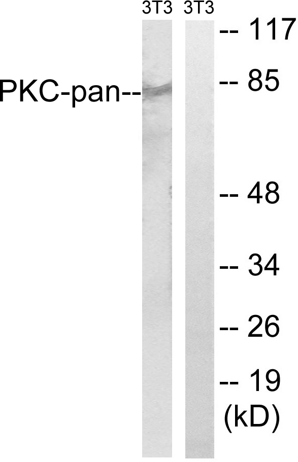
Anti-PKC-pan AntibodyA94967
ApplicationsImmunoFluorescence, Western Blot, ELISA, ImmunoHistoChemistry
ReactivityHuman, Mouse, Rat
- SizePrice
Product group Antibodies

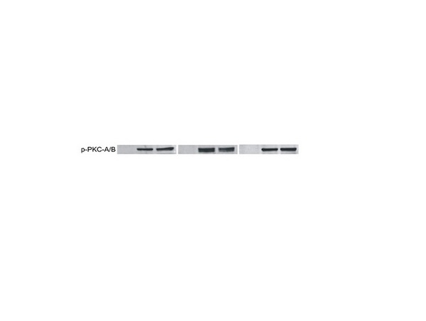
PKC alpha (Phospho-Tyr657) AntibodyABX012471
ApplicationsWestern Blot, ELISA
- SizePrice

Product group Antibodies
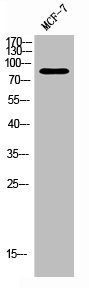
Anti-PKC Alpha/PRKCA Antibody Picoband(r)A00743-1-CARRIER-FREE
ApplicationsFlow Cytometry, Western Blot, ELISA
ReactivityHuman, Mouse, Rat
TargetPRKCA
- SizePrice
Product group Antibodies
Anti-PRKCA Antibody144-00267
ApplicationsImmunoFluorescence, Western Blot, ImmunoHistoChemistry
ReactivityHuman
TargetPRKCA
- SizePrice
Product group Antibodies
References
ApplicationsImmunoFluorescence, Western Blot, ELISA, ImmunoCytoChemistry, ImmunoHistoChemistry, ImmunoHistoChemistry Frozen, ImmunoHistoChemistry Paraffin
ReactivityBovine, Canine, Chicken, Equine, Human, Mouse, Rat
TargetPRKCA
- SizePrice
Product group Antibodies
ApplicationsImmunoFluorescence, Western Blot, ELISA, ImmunoHistoChemistry
ReactivityHuman, Mouse, Rat
TargetPRKCA
- SizePrice
Product group Antibodies
Prkca Polyclonal AntibodyCAC09189
ApplicationsImmunoFluorescence, ELISA, ImmunoHistoChemistry
TargetPRKCA
- SizePrice
Product group Antibodies

PRKCA / PKC-Alpha AntibodyLS-C402927
ApplicationsWestern Blot, ELISA
ReactivityHuman, Mouse, Rat
TargetPRKCA
- SizePrice