


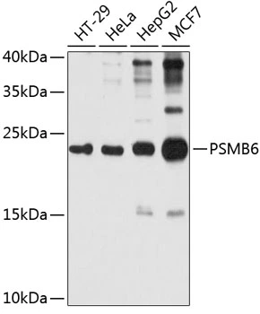
Figure 1. Western blot analysis of Proteasome 20S Beta 6/PSMB6 using anti-Proteasome 20S Beta 6/PSMB6 antibody (A07705-1). Electrophoresis was performed on a 5-20% SDS-PAGE gel at 70V (Stacking gel) / 90V (Resolving gel) for 2-3 hours. The sample well of each lane was loaded with 30 ug of sample under reducing conditions. Lane 1: human Caco-2 whole cell lysates, Lane 2: human Hela whole cell lysates, Lane 3: monkey COS-7 whole cell lysates, Lane 4: human A549 whole cell lysates, Lane 5: human Jurkat whole cell lysates, Lane 6: human MCF-7 whole cell lysates, Lane 7: rat liver tissue lysates, Lane 8: rat brain tissue lysates, Lane 9: rat lung tissue lysates, Lane 10: rat testis tissue lysates, Lane 11: mouse liver tissue lysates, Lane 12: mouse brain tissue lysates, Lane 13: mouse lung tissue lysates, Lane 14: mouse testis tissue lysates. After electrophoresis, proteins were transferred to a nitrocellulose membrane at 150 mA for 50-90 minutes. Blocked the membrane with 5% non-fat milk/TBS for 1.5 hour at RT. The membrane was incubated with rabbit anti-Proteasome 20S Beta 6/PSMB6 antigen affinity purified polyclonal antibody (Catalog # A07705-1) at 0.5 microg/mL overnight at 4°C, then washed with TBS-0.1%Tween 3 times with 5 minutes each and probed with a goat anti-rabbit IgG-HRP secondary antibody at a dilution of 1:5000 for 1.5 hour at RT. The signal is developed using an Enhanced Chemiluminescent detection (ECL) kit (Catalog # EK1002) with Tanon 5200 system. A specific band was detected for Proteasome 20S Beta 6/PSMB6 at approximately 22 kDa. The expected band size for Proteasome 20S Beta 6/PSMB6 is at 22 kDa.
. Overlay histogram showing A431 cells stained with A07705-1 (Blue line). To facilitate intracellular staining, cells were fixed with 4% paraformaldehyde and permeabilized with permeabilization buffer. The cells were blocked with 10% normal goat serum. And then incubated with rabbit anti-Proteasome 20S Beta 6/PSMB6 Antibody (A07705-1, 1 microg/1x106 cells) for 30 min at 20°C. DyLight®488 conjugated goat anti-rabbit IgG (BA1127, 5-10 microg/1x106 cells) was used as secondary antibody for 30 minutes at 20°C. Isotype control antibody (Green line) was rabbit IgG (1 microg/1x106) used under the same conditions. Unlabelled sample without incubation with primary antibody and secondary antibody (Red line) was used as a blank control.")
Figure 1. Western blot analysis of Proteasome 20S Beta 6/PSMB6 using anti-Proteasome 20S Beta 6/PSMB6 antibody (A07705-1). Electrophoresis was performed on a 5-20% SDS-PAGE gel at 70V (Stacking gel) / 90V (Resolving gel) for 2-3 hours. The sample well of each lane was loaded with 30 ug of sample under reducing conditions. Lane 1: human Caco-2 whole cell lysates, Lane 2: human Hela whole cell lysates, Lane 3: monkey COS-7 whole cell lysates, Lane 4: human A549 whole cell lysates, Lane 5: human Jurkat whole cell lysates, Lane 6: human MCF-7 whole cell lysates, Lane 7: rat liver tissue lysates, Lane 8: rat brain tissue lysates, Lane 9: rat lung tissue lysates, Lane 10: rat testis tissue lysates, Lane 11: mouse liver tissue lysates, Lane 12: mouse brain tissue lysates, Lane 13: mouse lung tissue lysates, Lane 14: mouse testis tissue lysates. After electrophoresis, proteins were transferred to a nitrocellulose membrane at 150 mA for 50-90 minutes. Blocked the membrane with 5% non-fat milk/TBS for 1.5 hour at RT. The membrane was incubated with rabbit anti-Proteasome 20S Beta 6/PSMB6 antigen affinity purified polyclonal antibody (Catalog # A07705-1) at 0.5 microg/mL overnight at 4°C, then washed with TBS-0.1%Tween 3 times with 5 minutes each and probed with a goat anti-rabbit IgG-HRP secondary antibody at a dilution of 1:5000 for 1.5 hour at RT. The signal is developed using an Enhanced Chemiluminescent detection (ECL) kit (Catalog # EK1002) with Tanon 5200 system. A specific band was detected for Proteasome 20S Beta 6/PSMB6 at approximately 22 kDa. The expected band size for Proteasome 20S Beta 6/PSMB6 is at 22 kDa.
Anti-Proteasome 20S beta 6/PSMB6 Antibody Picoband(r)
A07705-1-CARRIER-FREE
ApplicationsFlow Cytometry, Western Blot, ELISA
Product group Antibodies
ReactivityHuman, Monkey, Mouse, Rat
TargetPSMB6
Overview
- SupplierBoster Bio
- Product NameAnti-Proteasome 20S beta 6/PSMB6 Antibody Picoband(r)
- Delivery Days Customer9
- ApplicationsFlow Cytometry, Western Blot, ELISA
- CertificationResearch Use Only
- ClonalityPolyclonal
- Concentration500 ug/ml
- Gene ID5694
- Target namePSMB6
- Target descriptionproteasome 20S subunit beta 6
- Target synonymsDELTA, LMPY, proteasome subunit beta type-6, PSY large multifunctional protease Y, beta-1, macropain delta chain, multicatalytic endopeptidase complex delta chain, proteasome (prosome, macropain) subunit, beta type, 6, proteasome catalytic subunit 1, proteasome delta chain, proteasome subunit Y, proteasome subunit beta 6, proteasome subunit beta-1, proteasome subunit beta1, proteasome subunit delta
- HostRabbit
- IsotypeIgG
- Protein IDP28072
- Protein NameProteasome subunit beta type-6
- Scientific DescriptionBoster Bio Anti-Proteasome 20S beta 6/PSMB6 Antibody Picoband® catalog # A07705-1. Tested in ELISA, Flow Cytometry, WB applications. This antibody reacts with Human, Mouse, Rat. The brand Picoband indicates this is a premium antibody that guarantees superior quality, high affinity, and strong signals with minimal background in Western blot applications. Only our best-performing antibodies are designated as Picoband, ensuring unmatched performance.
- ReactivityHuman, Monkey, Mouse, Rat
- Storage Instruction-20°C,2°C to 8°C
- UNSPSC12352203
Related products
Product group Antibodies
Anti-PSMB6 Antibody144-04053
ApplicationsWestern Blot, ImmunoHistoChemistry
ReactivityHuman, Mouse, Rat
TargetPSMB6
- SizePrice
Product group Antibodies

PSMB6 AntibodyLS-C667981
ApplicationsWestern Blot
ReactivityHuman
TargetPSMB6
- SizePrice
Product group Antibodies
ApplicationsWestern Blot, ImmunoHistoChemistry
TargetPSMB6
- SizePrice
Product group Antibodies
PSMB6 AntibodyCSB-PA018884GA01HU
ApplicationsWestern Blot, ELISA, ImmunoHistoChemistry
ReactivityHuman, Mouse, Rat
TargetPSMB6
- SizePrice

Product group Antibodies
Anti-PSMB6 AntibodyHPA023312
ApplicationsWestern Blot, ImmunoHistoChemistry
ReactivityHuman
TargetPSMB6
- SizePrice
Product group Antibodies
PSMB6 antibodyGTX66015
ApplicationsImmunoFluorescence, Western Blot, ImmunoCytoChemistry
ReactivityHuman
TargetPSMB6
- SizePrice
Product group Antibodies
Anti-PSMB6 AntibodyCAB4053
ApplicationsImmunoFluorescence, Western Blot, ELISA, ImmunoCytoChemistry, ImmunoHistoChemistry, ImmunoHistoChemistry Paraffin
ReactivityHuman
TargetPSMB6
- SizePrice