


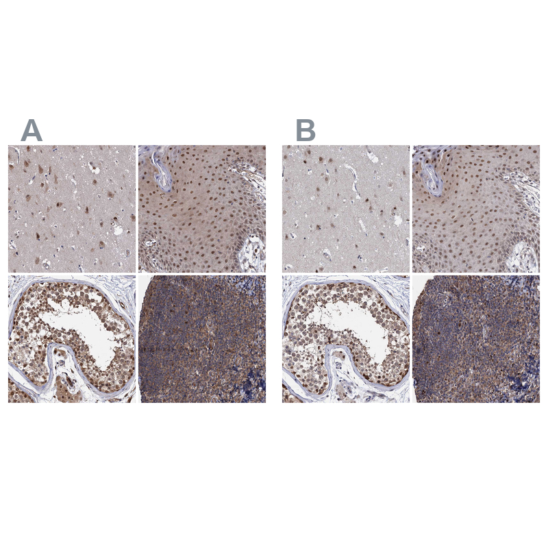
Immunohistochemical staining of human cerebral cortex, squamous epithelia, testis and tonsil using Anti-PSMD4 antibody HPA038807 (A) shows similar protein distribution across tissues to independent antibody HPA039252 (B).
 and PSMD4 over-expression lysate (Co-expressed with a C-terminal myc-DDK tag (~3.1 kDa) in mammalian HEK293T cells, LY400997).")
Immunohistochemical staining of human cerebral cortex, squamous epithelia, testis and tonsil using Anti-PSMD4 antibody HPA038807 (A) shows similar protein distribution across tissues to independent antibody HPA039252 (B).
Anti-PSMD4 Antibody
HPA038807
ApplicationsWestern Blot, ImmunoCytoChemistry, ImmunoHistoChemistry
Product group Antibodies
ReactivityHuman
TargetPSMD4
Overview
- SupplierAtlas Antibodies
- Product NameAnti-PSMD4 Antibody
- Delivery Days Customer4
- ApplicationsWestern Blot, ImmunoCytoChemistry, ImmunoHistoChemistry
- CertificationResearch Use Only
- ClonalityPolyclonal
- ConjugateUnconjugated
- Gene ID5710
- Target namePSMD4
- Target descriptionproteasome 26S subunit ubiquitin receptor, non-ATPase 4
- Target synonymsAF, AF-1, ASF, MCB1, Rpn10, S5A, pUB-R5, 26S proteasome non-ATPase regulatory subunit 4, 26S proteasome regulatory subunit S5A, S5a/antisecretory factor protein, angiocidin, antisecretory factor 1, multiubiquitin chain-binding protein, proteasome (prosome, macropain) 26S subunit, non-ATPase, 4, proteasome 26S subunit, non-ATPase 4
- HostRabbit
- IsotypeIgG
- Protein IDP55036
- Protein Name26S proteasome non-ATPase regulatory subunit 4
- Scientific DescriptionRecombinant Protein Epitope Signature Tag (PrEST) antigen sequence
- ReactivityHuman
- Storage Instruction-20°C,2°C to 8°C
- UNSPSC41116161
Datasheet
MSDS
Related products
Product group Antibodies
Anti-PSMD4 AntibodyA29142
ApplicationsWestern Blot, ImmunoHistoChemistry
ReactivityHuman, Mouse, Rat
- SizePrice

Product group Antibodies
Anti-PSMD4 Antibody Picoband(r)A03544-1-CARRIER-FREE
ApplicationsFlow Cytometry, ImmunoFluorescence, Western Blot, ELISA, ImmunoCytoChemistry, ImmunoHistoChemistry
ReactivityHuman, Mouse, Rat
TargetPSMD4
- SizePrice
Product group Antibodies
Anti-PSMD4 Antibody144-01061
ApplicationsWestern Blot
ReactivityHuman, Monkey, Mouse
TargetPSMD4
- SizePrice
Product group Antibodies
ApplicationsImmunoFluorescence, Western Blot, ELISA, ImmunoCytoChemistry, ImmunoHistoChemistry, ImmunoHistoChemistry Frozen, ImmunoHistoChemistry Paraffin
ReactivityBovine, Canine, Chicken, Equine, Human, Mouse, Rabbit, Rat, Sheep
TargetPSMD4
- SizePrice
Product group Antibodies
PSMD4 Polyclonal AntibodyCAC15722
ApplicationsWestern Blot, ELISA, ImmunoHistoChemistry
TargetPSMD4
- SizePrice
Product group Antibodies
PSMD4 AntibodyCSB-PA018908HA01HU
ApplicationsWestern Blot, ELISA, ImmunoHistoChemistry
ReactivityHuman
TargetPSMD4
- SizePrice
Product group Antibodies

PSMD4 / RPN10 AntibodyLS-C331231
ApplicationsWestern Blot, ImmunoHistoChemistry
ReactivityHuman, Monkey, Mouse
TargetPSMD4
- SizePrice
Product group Antibodies
Anti-PSMD4 AntibodyHPA039252
ApplicationsImmunoCytoChemistry, ImmunoHistoChemistry
ReactivityHuman
TargetPSMD4
- SizePrice