

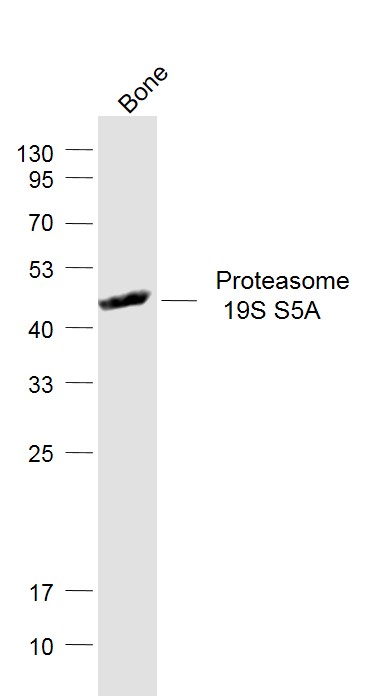
Mouse bone lysates probed with Proteasome 19S S5A Polyclonal Antibody, Unconjugated (bs-8322R) at 1:1000 dilution and 4˚C overnight incubation. Followed by conjugated secondary antibody incubation at 1:20000 for 60 min at 37˚C.
Mouse bone lysates probed with Proteasome 19S S5A Polyclonal Antibody, Unconjugated (bs-8322R) at 1:1000 dilution and 4˚C overnight incubation. Followed by conjugated secondary antibody incubation at 1:20000 for 60 min at 37˚C.
Proteasome 19S S5A Polyclonal Antibody
BS-8322R
ApplicationsImmunoFluorescence, Western Blot, ELISA, ImmunoCytoChemistry, ImmunoHistoChemistry, ImmunoHistoChemistry Frozen, ImmunoHistoChemistry Paraffin
Product group Antibodies
ReactivityBovine, Canine, Chicken, Equine, Human, Mouse, Rabbit, Rat, Sheep
TargetPSMD4
Overview
- SupplierBioss
- Product NameProteasome 19S S5A Polyclonal Antibody
- Delivery Days Customer16
- ApplicationsImmunoFluorescence, Western Blot, ELISA, ImmunoCytoChemistry, ImmunoHistoChemistry, ImmunoHistoChemistry Frozen, ImmunoHistoChemistry Paraffin
- Applications SupplierWB(1:300-5000), ELISA(1:500-1000), IHC-P(1:200-400), IHC-F(1:100-500), IF(IHC-P)(1:50-200), IF(IHC-F)(1:50-200), IF(ICC)(1:50-200)
- CertificationResearch Use Only
- ClonalityPolyclonal
- Concentration1 ug/ul
- ConjugateUnconjugated
- Gene ID5710
- Target namePSMD4
- Target descriptionproteasome 26S subunit ubiquitin receptor, non-ATPase 4
- Target synonymsAF, AF-1, ASF, MCB1, Rpn10, S5A, pUB-R5, 26S proteasome non-ATPase regulatory subunit 4, 26S proteasome regulatory subunit S5A, S5a/antisecretory factor protein, angiocidin, antisecretory factor 1, multiubiquitin chain-binding protein, proteasome (prosome, macropain) 26S subunit, non-ATPase, 4, proteasome 26S subunit, non-ATPase 4
- HostRabbit
- IsotypeIgG
- Protein IDP55036
- Protein Name26S proteasome non-ATPase regulatory subunit 4
- ReactivityBovine, Canine, Chicken, Equine, Human, Mouse, Rabbit, Rat, Sheep
- Storage Instruction-20°C
- UNSPSC41116161
Datasheet
Related products
Product group Antibodies
Anti-PSMD4 AntibodyA29142
ApplicationsWestern Blot, ImmunoHistoChemistry
ReactivityHuman, Mouse, Rat
- SizePrice

Product group Antibodies
Anti-PSMD4 Antibody Picoband(r)A03544-1-CARRIER-FREE
ApplicationsFlow Cytometry, ImmunoFluorescence, Western Blot, ELISA, ImmunoCytoChemistry, ImmunoHistoChemistry
ReactivityHuman, Mouse, Rat
TargetPSMD4
- SizePrice
Product group Antibodies
Anti-PSMD4 Antibody144-01061
ApplicationsWestern Blot
ReactivityHuman, Monkey, Mouse
TargetPSMD4
- SizePrice
Product group Antibodies
PSMD4 Polyclonal AntibodyCAC15722
ApplicationsWestern Blot, ELISA, ImmunoHistoChemistry
TargetPSMD4
- SizePrice
Product group Antibodies
PSMD4 AntibodyCSB-PA018908HA01HU
ApplicationsWestern Blot, ELISA, ImmunoHistoChemistry
ReactivityHuman
TargetPSMD4
- SizePrice
Product group Antibodies

PSMD4 / RPN10 AntibodyLS-C331231
ApplicationsWestern Blot, ImmunoHistoChemistry
ReactivityHuman, Monkey, Mouse
TargetPSMD4
- SizePrice
![PSMD4 antibody [N1C2] detects PSMD4 protein at cytoplasm and nucleus by immunofluorescent analysis. Sample: SK-N-SH cells were fixed in 4% paraformaldehyde at RT for 15 min. Green: PSMD4 protein stained by PSMD4 antibody [N1C2] (GTX114678) diluted at 1:500. Blue: Hoechst 33342 staining.](https://www.genetex.com/upload/website/prouct_img/normal/GTX114678/GTX114678_40212_IFA_w_23060518_950.webp)
Product group Antibodies
PSMD4 antibody [N1C2]GTX114678
ApplicationsImmunoFluorescence, Western Blot, ImmunoCytoChemistry
ReactivityHuman
TargetPSMD4
- SizePrice

Product group Antibodies
Anti-PSMD4 AntibodyHPA038807
ApplicationsWestern Blot, ImmunoCytoChemistry, ImmunoHistoChemistry
ReactivityHuman
TargetPSMD4
- SizePrice
Product group Antibodies
Anti-CYP7A1 AntibodyCAB10615
ApplicationsImmunoFluorescence, Western Blot, ELISA, ImmunoCytoChemistry
ReactivityHuman
TargetPSMD4
- SizePrice