

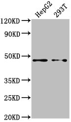
Western Blot Positive WB detected in: HepG2 whole cell lysate, 293T whole cell lysate All lanes: PSMD4 antibody at 1:2000 Secondary Goat polyclonal to rabbit IgG at 1/50000 dilution Predicted band size: 41, 29 kDa Observed band size: 41 kDa
. Section was blocked with 10% normal goat serum 30min at RT. Then primary antibody (1% BSA) was incubated at 4°C overnight. The primary is detected by a biotinylated secondary antibody and visualized using an HRP conjugated SP system.")
. Section was blocked with 10% normal goat serum 30min at RT. Then primary antibody (1% BSA) was incubated at 4°C overnight. The primary is detected by a biotinylated secondary antibody and visualized using an HRP conjugated SP system.")
Western Blot Positive WB detected in: HepG2 whole cell lysate, 293T whole cell lysate All lanes: PSMD4 antibody at 1:2000 Secondary Goat polyclonal to rabbit IgG at 1/50000 dilution Predicted band size: 41, 29 kDa Observed band size: 41 kDa
PSMD4 Antibody
CSB-PA018908HA01HU
ApplicationsWestern Blot, ELISA, ImmunoHistoChemistry
Product group Antibodies
ReactivityHuman
TargetPSMD4
Overview
- SupplierCusabio
- Product NamePSMD4 Antibody
- Delivery Days Customer20
- ApplicationsWestern Blot, ELISA, ImmunoHistoChemistry
- CertificationResearch Use Only
- ClonalityPolyclonal
- ConjugateUnconjugated
- Gene ID5710
- Target namePSMD4
- Target descriptionproteasome 26S subunit ubiquitin receptor, non-ATPase 4
- Target synonymsAF, AF-1, ASF, MCB1, Rpn10, S5A, pUB-R5, 26S proteasome non-ATPase regulatory subunit 4, 26S proteasome regulatory subunit S5A, S5a/antisecretory factor protein, angiocidin, antisecretory factor 1, multiubiquitin chain-binding protein, proteasome (prosome, macropain) 26S subunit, non-ATPase, 4, proteasome 26S subunit, non-ATPase 4
- HostRabbit
- IsotypeIgG
- Protein IDP55036
- Protein Name26S proteasome non-ATPase regulatory subunit 4
- Scientific DescriptionComponent of the 26S proteasome, a multiprotein complex involved in the ATP-dependent degradation of ubiquitinated proteins. This complex plays a key role in the maintenance of protein homeostasis by removing misfolded or damaged proteins, which could impair cellular functions, and by removing proteins whose functions are no longer required. Therefore, the proteasome participates in numerous cellular processes, including cell cycle progression, apoptosis, or DNA damage repair. PSMD4 acts as an ubiquitin receptor subunit through ubiquitin-interacting motifs and selects ubiquitin-conjugates for destruction. Displays a preferred selectivity for longer polyubiquitin chains.
- ReactivityHuman
- Storage Instruction-20°C or -80°C
- UNSPSC41116161
Related products
Product group Antibodies
Anti-PSMD4 AntibodyA29142
ApplicationsWestern Blot, ImmunoHistoChemistry
ReactivityHuman, Mouse, Rat
- SizePrice

Product group Antibodies
Anti-PSMD4 Antibody Picoband(r)A03544-1-CARRIER-FREE
ApplicationsFlow Cytometry, ImmunoFluorescence, Western Blot, ELISA, ImmunoCytoChemistry, ImmunoHistoChemistry
ReactivityHuman, Mouse, Rat
TargetPSMD4
- SizePrice
Product group Antibodies
Anti-PSMD4 Antibody144-01061
ApplicationsWestern Blot
ReactivityHuman, Monkey, Mouse
TargetPSMD4
- SizePrice
Product group Antibodies
ApplicationsImmunoFluorescence, Western Blot, ELISA, ImmunoCytoChemistry, ImmunoHistoChemistry, ImmunoHistoChemistry Frozen, ImmunoHistoChemistry Paraffin
ReactivityBovine, Canine, Chicken, Equine, Human, Mouse, Rabbit, Rat, Sheep
TargetPSMD4
- SizePrice
Product group Antibodies
PSMD4 Polyclonal AntibodyCAC15722
ApplicationsWestern Blot, ELISA, ImmunoHistoChemistry
TargetPSMD4
- SizePrice
Product group Antibodies

PSMD4 / RPN10 AntibodyLS-C331231
ApplicationsWestern Blot, ImmunoHistoChemistry
ReactivityHuman, Monkey, Mouse
TargetPSMD4
- SizePrice
![PSMD4 antibody [N1C2] detects PSMD4 protein at cytoplasm and nucleus by immunofluorescent analysis. Sample: SK-N-SH cells were fixed in 4% paraformaldehyde at RT for 15 min. Green: PSMD4 protein stained by PSMD4 antibody [N1C2] (GTX114678) diluted at 1:500. Blue: Hoechst 33342 staining.](https://www.genetex.com/upload/website/prouct_img/normal/GTX114678/GTX114678_40212_IFA_w_23060518_950.webp)
Product group Antibodies
PSMD4 antibody [N1C2]GTX114678
ApplicationsImmunoFluorescence, Western Blot, ImmunoCytoChemistry
ReactivityHuman
TargetPSMD4
- SizePrice

Product group Antibodies
Anti-PSMD4 AntibodyHPA038807
ApplicationsWestern Blot, ImmunoCytoChemistry, ImmunoHistoChemistry
ReactivityHuman
TargetPSMD4
- SizePrice
Product group Antibodies
Anti-CYP7A1 AntibodyCAB10615
ApplicationsImmunoFluorescence, Western Blot, ELISA, ImmunoCytoChemistry
ReactivityHuman
TargetPSMD4
- SizePrice