


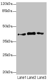
Figure 1. Western blot analysis of PTGES2/Gbf1 using anti-PTGES2/Gbf1 antibody (A06706-2). Electrophoresis was performed on a 5-20% SDS-PAGE gel at 70V (Stacking gel) / 90V (Resolving gel) for 2-3 hours. The sample well of each lane was loaded with 30 ug of sample under reducing conditions. Lane 1: human Hela whole cell lysates, Lane 2: human Hacat whole cell lysates, Lane 3: human MCF-7 whole cell lysates, Lane 4: human A549 whole cell lysates, Lane 5: rat brain tissue lysates, Lane 6: mouse brain tissue lysates. After electrophoresis, proteins were transferred to a nitrocellulose membrane at 150 mA for 50-90 minutes. Blocked the membrane with 5% non-fat milk/TBS for 1.5 hour at RT. The membrane was incubated with rabbit anti-PTGES2/Gbf1 antigen affinity purified polyclonal antibody (Catalog # A06706-2) at 0.5 microg/mL overnight at 4°C, then washed with TBS-0.1%Tween 3 times with 5 minutes each and probed with a goat anti-rabbit IgG-HRP secondary antibody at a dilution of 1:5000 for 1.5 hour at RT. The signal is developed using an Enhanced Chemiluminescent detection (ECL) kit (Catalog # EK1002) with Tanon 5200 system. A specific band was detected for PTGES2/Gbf1 at approximately 42 kDa. The expected band size for PTGES2/Gbf1 is at 42 kDa.
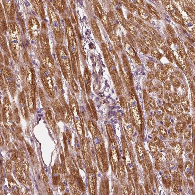
. PTGES2/Gbf1 was detected in a paraffin-embedded section of human breast cancer tissue. Heat mediated antigen retrieval was performed in EDTA buffer (pH 8.0, epitope retrieval solution). The tissue section was blocked with 10% goat serum. The tissue section was then incubated with 2 microg/ml rabbit anti-PTGES2/Gbf1 Antibody (A06706-2) overnight at 4°C. Biotinylated goat anti-rabbit IgG was used as secondary antibody and incubated for 30 minutes at 37°C. The tissue section was developed using Strepavidin-Biotin-Complex (SABC) (Catalog # SA1022) with DAB as the chromogen.")
. PTGES2/Gbf1 was detected in a paraffin-embedded section of human ovarian cancer tissue. Heat mediated antigen retrieval was performed in EDTA buffer (pH 8.0, epitope retrieval solution). The tissue section was blocked with 10% goat serum. The tissue section was then incubated with 2 microg/ml rabbit anti-PTGES2/Gbf1 Antibody (A06706-2) overnight at 4°C. Biotinylated goat anti-rabbit IgG was used as secondary antibody and incubated for 30 minutes at 37°C. The tissue section was developed using Strepavidin-Biotin-Complex (SABC) (Catalog # SA1022) with DAB as the chromogen.")
. PTGES2/Gbf1 was detected in an immunocytochemical section of Hela cells. Enzyme antigen retrieval was performed using IHC enzyme antigen retrieval reagent (AR0022) for 15 mins. The cells were blocked with 10% goat serum. And then incubated with 5 microg/mL rabbit anti-PTGES2/Gbf1 Antibody (A06706-2) overnight at 4°C. Cy3 Conjugated Goat Anti-Rabbit IgG (BA1032) was used as secondary antibody at 1:500 dilution and incubated for 30 minutes at 37°C. The section was counterstained with DAPI. Visualize using a fluorescence microscope and filter sets appropriate for the label used.")
. Overlay histogram showing MCF-7 cells stained with A06706-2 (Blue line). To facilitate intracellular staining, cells were fixed with 4% paraformaldehyde and permeabilized with permeabilization buffer. The cells were blocked with 10% normal goat serum. And then incubated with rabbit anti-PTGES2/Gbf1 Antibody (A06706-2, 1 microg/1x106 cells) for 30 min at 20°C. DyLight®488 conjugated goat anti-rabbit IgG (BA1127, 5-10 microg/1x106 cells) was used as secondary antibody for 30 minutes at 20°C. Isotype control antibody (Green line) was rabbit IgG (1 microg/1x106) used under the same conditions. Unlabelled sample (Red line) was also used as a control.")
Figure 1. Western blot analysis of PTGES2/Gbf1 using anti-PTGES2/Gbf1 antibody (A06706-2). Electrophoresis was performed on a 5-20% SDS-PAGE gel at 70V (Stacking gel) / 90V (Resolving gel) for 2-3 hours. The sample well of each lane was loaded with 30 ug of sample under reducing conditions. Lane 1: human Hela whole cell lysates, Lane 2: human Hacat whole cell lysates, Lane 3: human MCF-7 whole cell lysates, Lane 4: human A549 whole cell lysates, Lane 5: rat brain tissue lysates, Lane 6: mouse brain tissue lysates. After electrophoresis, proteins were transferred to a nitrocellulose membrane at 150 mA for 50-90 minutes. Blocked the membrane with 5% non-fat milk/TBS for 1.5 hour at RT. The membrane was incubated with rabbit anti-PTGES2/Gbf1 antigen affinity purified polyclonal antibody (Catalog # A06706-2) at 0.5 microg/mL overnight at 4°C, then washed with TBS-0.1%Tween 3 times with 5 minutes each and probed with a goat anti-rabbit IgG-HRP secondary antibody at a dilution of 1:5000 for 1.5 hour at RT. The signal is developed using an Enhanced Chemiluminescent detection (ECL) kit (Catalog # EK1002) with Tanon 5200 system. A specific band was detected for PTGES2/Gbf1 at approximately 42 kDa. The expected band size for PTGES2/Gbf1 is at 42 kDa.
Anti-PTGES2/Gbf1 Antibody Picoband(r)
A06706-2-CARRIER-FREE
ApplicationsFlow Cytometry, ImmunoFluorescence, Western Blot, ELISA, ImmunoCytoChemistry, ImmunoHistoChemistry
Product group Antibodies
ReactivityHuman, Mouse, Rat
TargetPTGES2
Overview
- SupplierBoster Bio
- Product NameAnti-PTGES2/Gbf1 Antibody Picoband(r)
- Delivery Days Customer9
- ApplicationsFlow Cytometry, ImmunoFluorescence, Western Blot, ELISA, ImmunoCytoChemistry, ImmunoHistoChemistry
- CertificationResearch Use Only
- ClonalityPolyclonal
- Concentration500 ug/ml
- Gene ID80142
- Target namePTGES2
- Target descriptionprostaglandin E synthase 2
- Target synonymsC9orf15, GBF-1, GBF1, PGES2, mPGES-2, prostaglandin E synthase 2, GATE-binding factor 1, gamma-interferon-activated transcriptional element-binding factor 1, mPGE synthase-2, membrane-associated prostaglandin E synthase 2, microsomal prostaglandin E synthase-2, prostaglandin-H(2) E-isomerase
- HostRabbit
- IsotypeIgG
- Protein IDQ9H7Z7
- Protein NameProstaglandin E synthase 2
- Scientific DescriptionBoster Bio Anti-PTGES2/Gbf1 Antibody Picoband® catalog # A06706-2. Tested in ELISA, Flow Cytometry, IF, IHC, ICC, WB applications. This antibody reacts with Human, Mouse, Rat. The brand Picoband indicates this is a premium antibody that guarantees superior quality, high affinity, and strong signals with minimal background in Western blot applications. Only our best-performing antibodies are designated as Picoband, ensuring unmatched performance.
- ReactivityHuman, Mouse, Rat
- Storage Instruction-20°C,2°C to 8°C
- UNSPSC12352203
Related products
Product group Antibodies
PTGES2 AntibodyCSB-PA018977ESR1HU
ApplicationsWestern Blot, ELISA, ImmunoHistoChemistry
ReactivityHuman
TargetPTGES2
- SizePrice
Product group Antibodies
Anti-PTGES2 AntibodyA82770
ApplicationsWestern Blot, ELISA, ImmunoHistoChemistry
ReactivityHuman
- SizePrice
Product group Antibodies

PTGES2 AntibodyLS-C748491
ApplicationsImmunoFluorescence, Western Blot
ReactivityHuman, Mouse, Rat
TargetPTGES2
- SizePrice
Product group Antibodies
Anti-PTGES2 AntibodyHPA020733
ApplicationsWestern Blot, ImmunoHistoChemistry
ReactivityHuman
TargetPTGES2
- SizePrice
Product group Antibodies

Goat anti-PTGES2 (aa292-306)EB12928
ApplicationsWestern Blot, ELISA, ImmunoHistoChemistry
ReactivityHuman
TargetPTGES2
- SizePrice
Product group Antibodies
ApplicationsFlow Cytometry
TargetPTGES2
- SizePrice
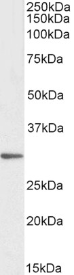
![Various whole cell extracts (30 μg) were separated by 10% SDS-PAGE, and the membrane was blotted with PTGES2 antibody [N2C3] (GTX112174) diluted at 1:1000. The HRP-conjugated anti-rabbit IgG antibody (GTX213110-01) was used to detect the primary antibody.](https://www.genetex.com/upload/website/prouct_img/normal/GTX112174/GTX112174_40107_20190913_WB_w_23060500_797.webp)
Product group Antibodies
PTGES2 antibody [N2C3]GTX112174
ApplicationsWestern Blot
ReactivityHuman
TargetPTGES2
- SizePrice
Product group Antibodies
Anti-PTGES2 Antibody144-07137
ApplicationsImmunoFluorescence, Western Blot, ImmunoHistoChemistry
ReactivityHuman, Mouse, Rat
TargetPTGES2
- SizePrice
Product group Antibodies
PTGES2 Polyclonal AntibodyBS-25192R
ApplicationsImmunoFluorescence, Western Blot, ImmunoHistoChemistry, ImmunoHistoChemistry Frozen, ImmunoHistoChemistry Paraffin
ReactivityBovine, Canine, Human, Mouse, Porcine, Rabbit, Rat
TargetPTGES2
- SizePrice