


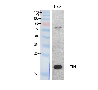
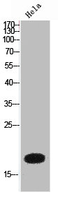
Figure 1. Western blot analysis of PTN using anti-PTN antibody (A01368-2). Electrophoresis was performed on a 5-20% SDS-PAGE gel at 70V (Stacking gel) / 90V (Resolving gel) for 2-3 hours. The sample well of each lane was loaded with 30 ug of sample under reducing conditions. Lane 1: human U251 whole cell lysates, Lane 2: rat brain tissue lysates, Lane 3: rat C6 whole cell lysates, Lane 4: mouse brain tissue cell lysates. After electrophoresis, proteins were transferred to a nitrocellulose membrane at 150 mA for 50-90 minutes. Blocked the membrane with 5% non-fat milk/TBS for 1.5 hour at RT. The membrane was incubated with rabbit anti-PTN antigen affinity purified polyclonal antibody (Catalog # A01368-2) at 0.5 microg/mL overnight at 4°C, then washed with TBS-0.1%Tween 3 times with 5 minutes each and probed with a goat anti-rabbit IgG-HRP secondary antibody at a dilution of 1:5000 for 1.5 hour at RT. The signal is developed using an Enhanced Chemiluminescent detection (ECL) kit (Catalog # EK1002) with Tanon 5200 system. A specific band was detected for PTN at approximately 19 kDa. The expected band size for PTN is at 19 kDa.
. PTN was detected in a paraffin-embedded section of human glioma tissue. Heat mediated antigen retrieval was performed in EDTA buffer (pH 8.0, epitope retrieval solution). The tissue section was blocked with 10% goat serum. The tissue section was then incubated with 2 microg/ml rabbit anti-PTN Antibody (A01368-2) overnight at 4°C. Peroxidase Conjugated Goat Anti-rabbit IgG was used as secondary antibody and incubated for 30 minutes at 37°C. The tissue section was developed using HRP Conjugated Rabbit IgG Super Vision Assay Kit (Catalog # SV0002) with DAB as the chromogen.")
. PTN was detected in a paraffin-embedded section of mouse brain tissue. Heat mediated antigen retrieval was performed in EDTA buffer (pH 8.0, epitope retrieval solution). The tissue section was blocked with 10% goat serum. The tissue section was then incubated with 2 microg/ml rabbit anti-PTN Antibody (A01368-2) overnight at 4°C. Peroxidase Conjugated Goat Anti-rabbit IgG was used as secondary antibody and incubated for 30 minutes at 37°C. The tissue section was developed using HRP Conjugated Rabbit IgG Super Vision Assay Kit (Catalog # SV0002) with DAB as the chromogen.")
. PTN was detected in a paraffin-embedded section of rat brain tissue. Heat mediated antigen retrieval was performed in EDTA buffer (pH 8.0, epitope retrieval solution). The tissue section was blocked with 10% goat serum. The tissue section was then incubated with 2 microg/ml rabbit anti-PTN Antibody (A01368-2) overnight at 4°C. Peroxidase Conjugated Goat Anti-rabbit IgG was used as secondary antibody and incubated for 30 minutes at 37°C. The tissue section was developed using HRP Conjugated Rabbit IgG Super Vision Assay Kit (Catalog # SV0002) with DAB as the chromogen.")
. Overlay histogram showing U20S cells stained with A01368-2 (Blue line). The cells were fixed with 4% paraformaldehyde and blocked with 10% normal goat serum. And then incubated with rabbit anti-PTN Antibody (A01368-2, 1 microg/1x106 cells) for 30 min at 20°C. DyLight®488 conjugated goat anti-rabbit IgG (BA1127, 5-10 microg/1x106 cells) was used as secondary antibody for 30 minutes at 20°C. Isotype control antibody (Green line) was rabbit IgG (1 microg/1x106) used under the same conditions. Unlabelled sample without incubation with primary antibody and secondary antibody (Red line) was used as a blank control.")
Figure 1. Western blot analysis of PTN using anti-PTN antibody (A01368-2). Electrophoresis was performed on a 5-20% SDS-PAGE gel at 70V (Stacking gel) / 90V (Resolving gel) for 2-3 hours. The sample well of each lane was loaded with 30 ug of sample under reducing conditions. Lane 1: human U251 whole cell lysates, Lane 2: rat brain tissue lysates, Lane 3: rat C6 whole cell lysates, Lane 4: mouse brain tissue cell lysates. After electrophoresis, proteins were transferred to a nitrocellulose membrane at 150 mA for 50-90 minutes. Blocked the membrane with 5% non-fat milk/TBS for 1.5 hour at RT. The membrane was incubated with rabbit anti-PTN antigen affinity purified polyclonal antibody (Catalog # A01368-2) at 0.5 microg/mL overnight at 4°C, then washed with TBS-0.1%Tween 3 times with 5 minutes each and probed with a goat anti-rabbit IgG-HRP secondary antibody at a dilution of 1:5000 for 1.5 hour at RT. The signal is developed using an Enhanced Chemiluminescent detection (ECL) kit (Catalog # EK1002) with Tanon 5200 system. A specific band was detected for PTN at approximately 19 kDa. The expected band size for PTN is at 19 kDa.
Anti-PTN Antibody Picoband(r)
A01368-2-FITC
ApplicationsFlow Cytometry, Western Blot, ELISA, ImmunoHistoChemistry
Product group Antibodies
ReactivityHuman, Mouse, Rat
TargetPTN
Overview
- SupplierBoster Bio
- Product NameAnti-PTN Antibody Picoband(r)
- Delivery Days Customer9
- ApplicationsFlow Cytometry, Western Blot, ELISA, ImmunoHistoChemistry
- CertificationResearch Use Only
- ClonalityPolyclonal
- Concentration500 ug/ml
- ConjugateFITC
- Gene ID5764
- Target namePTN
- Target descriptionpleiotrophin
- Target synonymsHARP, HB-GAM, HBBM, HBGF-8, HBGF8, HBNF, HBNF-1, NEGF1, OSF-1, pleiotrophin, heparin affin regulatory protein, heparin-binding brain mitogen, heparin-binding growth factor 8, heparin-binding growth-associated molecule, heparin-binding neurite outgrowth promoting factor, heparin-binding neurite outgrowth-promoting factor 1, osteoblast-specific factor 1, pleiotrophin (heparin binding growth factor 8, neurite growth-promoting factor 1)
- HostRabbit
- IsotypeIgG
- Protein IDP21246
- Protein NamePleiotrophin
- Scientific DescriptionBoster Bio Anti-PTN Antibody Picoband® catalog # A01368-2. Tested in ELISA, Flow Cytometry, IHC, WB applications. This antibody reacts with Human, Mouse, Rat. The brand Picoband indicates this is a premium antibody that guarantees superior quality, high affinity, and strong signals with minimal background in Western blot applications. Only our best-performing antibodies are designated as Picoband, ensuring unmatched performance.
- ReactivityHuman, Mouse, Rat
- Storage Instruction-20°C,2°C to 8°C
- UNSPSC12352203
Related products
Product group Antibodies
PTN Polyclonal Antibody, AbBy Fluor-350 ConjugatedBS-21928R-BF350
ApplicationsImmunoFluorescence, ImmunoCytoChemistry, ImmunoHistoChemistry, ImmunoHistoChemistry Frozen, ImmunoHistoChemistry Paraffin
ReactivityEquine, Human, Mouse, Rabbit, Rat
TargetPTN
- SizePrice
Product group Antibodies
ApplicationsImmunoPrecipitation, Western Blot, ImmunoCytoChemistry, ImmunoHistoChemistry
TargetPTN
- SizePrice
Product group Antibodies
Anti-PTN (N-term) Antibody102-27382
ApplicationsWestern Blot, ImmunoHistoChemistry, ImmunoHistoChemistry Paraffin
TargetPTN
- SizePrice
Product group Antibodies
Anti-PTN AntibodyAMAB92060
ApplicationsImmunoHistoChemistry
ReactivityHuman, Mouse
TargetPTN
- SizePrice
Product group Antibodies
Anti-PTN AntibodyA98457
ApplicationsWestern Blot, ELISA
ReactivityHuman, Mouse, Rat
- SizePrice
Product group Antibodies

ApplicationsWestern Blot, ELISA, ImmunoHistoChemistry
ReactivityHuman, Mouse, Rat
TargetPTN
- SizePrice
Product group Antibodies
Pleiotrophin antibodyGTX10849
ApplicationsWestern Blot, ELISA
ReactivityHuman
TargetPTN
- SizePrice
Product group Antibodies

PTN / Pleiotrophin AntibodyLS-C402762
ApplicationsWestern Blot, ELISA
ReactivityHuman, Mouse, Rat
TargetPTN
- SizePrice
Product group Antibodies
PTN AntibodyCSB-PA006030
ApplicationsWestern Blot, ELISA
ReactivityHuman, Mouse, Rat
TargetPTN
- SizePrice