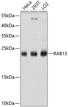
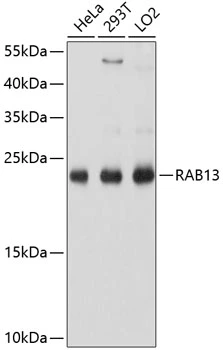
Bio-Connect
Anti-RAB13 Antibody

Anti-RAB13 Antibody

A8656
Antibodies.com
ApplicationsImmunoFluorescence, Western Blot, ImmunoCytoChemistry
Product group Antibodies
Sign in to order and to see your custom pricing.
Large volume orders?
Order with a bulk request

Overview

  • Supplier
    Antibodies.com
  • Product Name
    Anti-RAB13 Antibody
  • Delivery Days Customer
    7
  • Applications
    ImmunoFluorescence, Western Blot, ImmunoCytoChemistry
  • Certification
    Research Use Only
  • Clonality
    Polyclonal
  • Conjugate
    Unconjugated
  • Host
    Rabbit
  • Isotype
    IgG
  • Scientific Description
    Rabbit polyclonal antibody to RAB13.
  • UNSPSC
    12352203

Related products

WB analysis of various sample lysates using GTX64803 RAB13 antibody. Dilution : 1:1000 Loading : 25microg per lane
Product group Antibodies
GeneTex
ApplicationsImmunoFluorescence, Western Blot, ImmunoCytoChemistry
TargetRAB13
  • SizePrice
Immunohistochemical staining of human stomach shows moderate membranous positivity in glandular cells.
Product group Antibodies
Atlas Antibodies
ApplicationsWestern Blot, ImmunoCytoChemistry, ImmunoHistoChemistry
TargetRAB13
  • SizePrice
Product group Antibodies
RayBiotech
ApplicationsWestern Blot
TargetRAB13
  • SizePrice
Figure 1. Western blot analysis of RAB13 using anti-RAB13 antibody (A04339). Electrophoresis was performed on a 5-20% SDS-PAGE gel at 70V (Stacking gel) / 90V (Resolving gel) for 2-3 hours. The sample well of each lane was loaded with 30 ug of sample under reducing conditions. Lane 1: human Hela whole cell lysates, Lane 2: human Caco-2 whole cell lysates, Lane 3: human MCF-7 whole cell lysates, Lane 4: human 293T whole cell lysates. After electrophoresis, proteins were transferred to a nitrocellulose membrane at 150 mA for 50-90 minutes. Blocked the membrane with 5% non-fat milk/TBS for 1.5 hour at RT. The membrane was incubated with rabbit anti-RAB13 antigen affinity purified polyclonal antibody (Catalog # A04339) at 0.25 microg/mL overnight at 4°C, then washed with TBS-0.1%Tween 3 times with 5 minutes each and probed with a goat anti-rabbit IgG-HRP secondary antibody at a dilution of 1:5000 for 1.5 hour at RT. The signal is developed using an Enhanced Chemiluminescent detection (ECL) kit (Catalog # EK1002) with Tanon 5200 system. A specific band was detected for RAB13 at approximately 23 kDa. The expected band size for RAB13 is at 23 kDa.
Product group Antibodies
Boster Bio
ApplicationsFlow Cytometry, ImmunoFluorescence, Western Blot, ELISA, ImmunoCytoChemistry, ImmunoHistoChemistry
TargetRAB13
  • SizePrice
Lane 1: MCF-7; Lane 2: A431; Lane 3: A549; Lane 4: U-87MG cell lysate at 20 µg per lane, probed with bsm-51357M RAB13 (5C9) Monoclonal Antibody at 1:1000 dilution and 4℃ overnight incubation, followed by secondary antibody incubation for 60min at room temperature.
Product group Antibodies
Bioss Antibodies
ApplicationsWestern Blot
TargetRAB13
  • SizePrice
Product group Antibodies
MedChem Express
TargetRAB13
  • SizePrice
Product group Antibodies
LifeSpan BioSciences
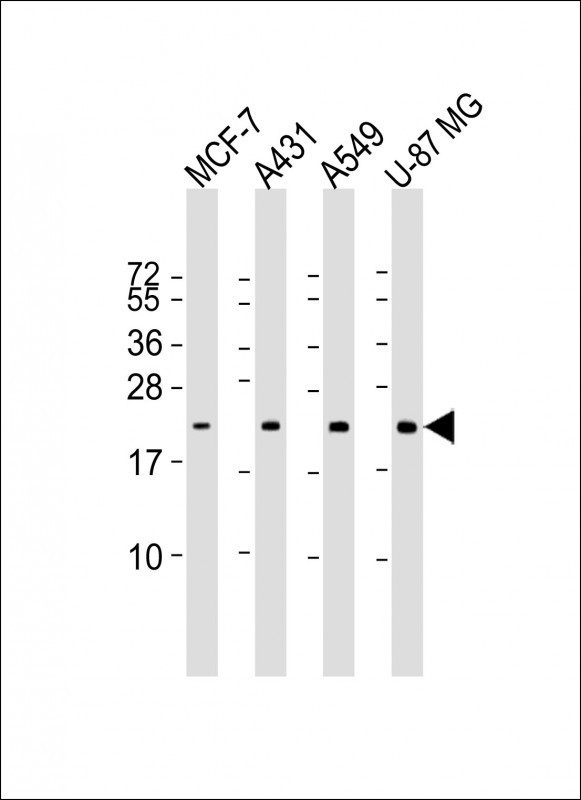
RAB13 AntibodyLS-C830922
ApplicationsELISA, ImmunoHistoChemistry
TargetRAB13
  • SizePrice