

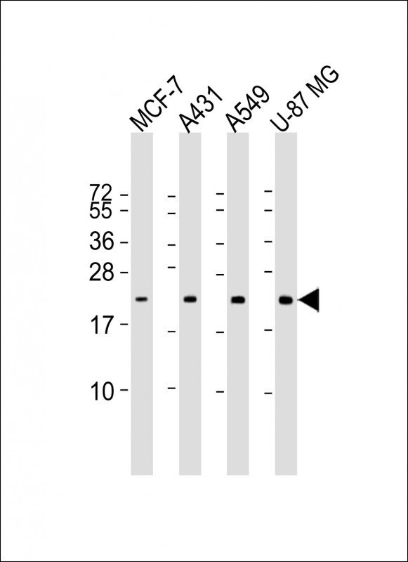
Lane 1: MCF-7; Lane 2: A431; Lane 3: A549; Lane 4: U-87MG cell lysate at 20 µg per lane, probed with bsm-51357M RAB13 (5C9) Monoclonal Antibody at 1:1000 dilution and 4℃ overnight incubation, followed by secondary antibody incubation for 60min at room temperature.
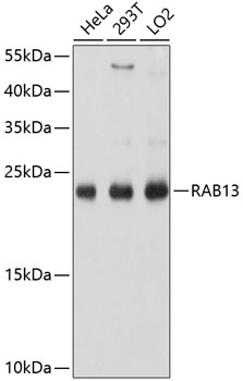
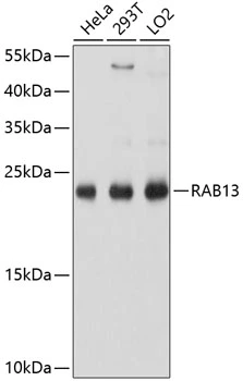
 Monoclonal Antibody, unconjugated (bsm-51357M) at 1:1000 overnight at 4°C followed by a conjugated secondary antibody for 60 minutes at 37°C.")
Lane 1: MCF-7; Lane 2: A431; Lane 3: A549; Lane 4: U-87MG cell lysate at 20 µg per lane, probed with bsm-51357M RAB13 (5C9) Monoclonal Antibody at 1:1000 dilution and 4℃ overnight incubation, followed by secondary antibody incubation for 60min at room temperature.
RAB13 (5C9) Monoclonal Antibody
BSM-51357M
ApplicationsWestern Blot
Product group Antibodies
ReactivityHuman
TargetRAB13
Overview
- SupplierBioss
- Product NameRAB13 (5C9) Monoclonal Antibody
- Delivery Days Customer16
- ApplicationsWestern Blot
- Applications SupplierWB(1:300-5000)
- CertificationResearch Use Only
- ClonalityMonoclonal
- Clone ID5C9
- Concentration0.5 ug/ml
- ConjugateUnconjugated
- Gene ID5872
- Target nameRAB13
- Target descriptionRAB13, member RAS oncogene family
- Target synonymsGIG4, ras-related protein Rab-13, RAS-associated protein RAB13, cell growth-inhibiting gene 4 protein, growth-inhibiting gene 4 protein
- HostMouse
- IsotypeIgG1
- Protein IDP51153
- Protein NameRas-related protein Rab-13
- ReactivityHuman
- Storage Instruction-20°C
- UNSPSC41116161
Datasheet
Related products
Product group Antibodies
Anti-RAB13 AntibodyA8656
ApplicationsImmunoFluorescence, Western Blot, ImmunoCytoChemistry
ReactivityHuman, Rat
- SizePrice
Product group Antibodies

RAB13 AntibodyLS-C830922
ApplicationsELISA, ImmunoHistoChemistry
ReactivityHuman, Mouse, Rat
TargetRAB13
- SizePrice

Product group Antibodies
Anti-RAB13 AntibodyHPA003996
ApplicationsWestern Blot, ImmunoCytoChemistry, ImmunoHistoChemistry
ReactivityHuman
TargetRAB13
- SizePrice

Product group Antibodies
Anti-RAB13 Antibody Picoband(r)A04339-CARRIER-FREE
ApplicationsFlow Cytometry, ImmunoFluorescence, Western Blot, ELISA, ImmunoCytoChemistry, ImmunoHistoChemistry
ReactivityHuman, Rat
TargetRAB13
- SizePrice
Product group Antibodies
RAB13 antibodyGTX64803
ApplicationsImmunoFluorescence, Western Blot, ImmunoCytoChemistry
ReactivityHuman
TargetRAB13
- SizePrice
Product group Antibodies
Anti-RAB13 Antibody144-10571
ApplicationsWestern Blot
ReactivityHuman, Mouse, Rat
TargetRAB13
- SizePrice