


Immunohistochemical staining of human stomach shows moderate membranous positivity in glandular cells.

Immunohistochemical staining of human stomach shows moderate membranous positivity in glandular cells.
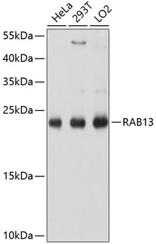
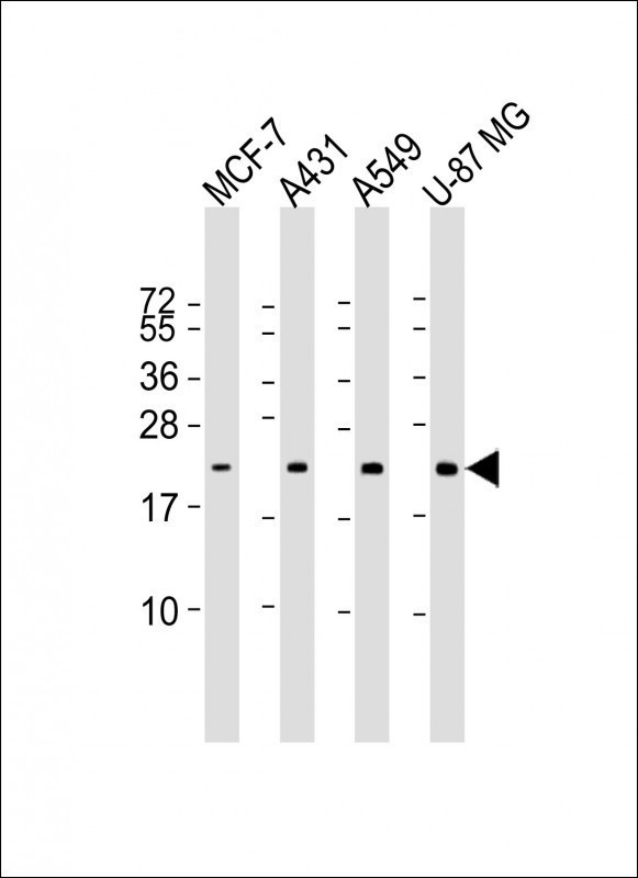
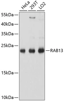
Anti-RAB13 Antibody
HPA003996
ApplicationsWestern Blot, ImmunoCytoChemistry, ImmunoHistoChemistry
Product group Antibodies
ReactivityHuman
TargetRAB13
Overview
- SupplierAtlas Antibodies
- Product NameAnti-RAB13 Antibody
- Delivery Days Customer4
- ApplicationsWestern Blot, ImmunoCytoChemistry, ImmunoHistoChemistry
- CertificationResearch Use Only
- ClonalityPolyclonal
- ConjugateUnconjugated
- Gene ID5872
- Target nameRAB13
- Target descriptionRAB13, member RAS oncogene family
- Target synonymsGIG4, ras-related protein Rab-13, RAS-associated protein RAB13, cell growth-inhibiting gene 4 protein, growth-inhibiting gene 4 protein
- HostRabbit
- IsotypeIgG
- Protein IDP51153
- Protein NameRas-related protein Rab-13
- Scientific DescriptionRecombinant Protein Epitope Signature Tag (PrEST) antigen sequence
- ReactivityHuman
- Storage Instruction-20°C,2°C to 8°C
- UNSPSC41116161
Datasheet
MSDS
Related products
Product group Antibodies
Anti-RAB13 AntibodyA8656
ApplicationsImmunoFluorescence, Western Blot, ImmunoCytoChemistry
ReactivityHuman, Rat
- SizePrice
Product group Antibodies

RAB13 AntibodyLS-C830922
ApplicationsELISA, ImmunoHistoChemistry
ReactivityHuman, Mouse, Rat
TargetRAB13
- SizePrice

Product group Antibodies
Anti-RAB13 Antibody Picoband(r)A04339-CARRIER-FREE
ApplicationsFlow Cytometry, ImmunoFluorescence, Western Blot, ELISA, ImmunoCytoChemistry, ImmunoHistoChemistry
ReactivityHuman, Rat
TargetRAB13
- SizePrice
Product group Antibodies
RAB13 (5C9) Monoclonal AntibodyBSM-51357M
ApplicationsWestern Blot
ReactivityHuman
TargetRAB13
- SizePrice
Product group Antibodies
RAB13 antibodyGTX64803
ApplicationsImmunoFluorescence, Western Blot, ImmunoCytoChemistry
ReactivityHuman
TargetRAB13
- SizePrice
Product group Antibodies
Anti-RAB13 Antibody144-10571
ApplicationsWestern Blot
ReactivityHuman, Mouse, Rat
TargetRAB13
- SizePrice