

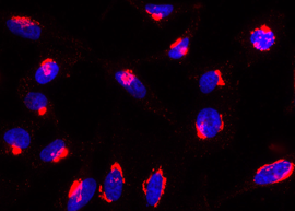
Rab6-GTP is detected by immunocytochemistry using anti-Rab6-GTP, mAb (AA2) (Prod. No. AG-27B-0004). Method: HeLa cells are grown in standard culture conditions, fixed with paraformaldehyde (3%), permeablized in PBS+ BSA 0.2 % + Sap
Rab6-GTP is detected by immunocytochemistry using anti-Rab6-GTP, mAb (AA2) (Prod. No. AG-27B-0004). Method: HeLa cells are grown in standard culture conditions, fixed with paraformaldehyde (3%), permeablized in PBS+ BSA 0.2 % + Sap
anti-Rab6-GTP, mAb (rec.) (AA2)
AG-27B-0004
ApplicationsImmunoCytoChemistry
Product group Antibodies
ReactivityDrosophila, Human, Mouse
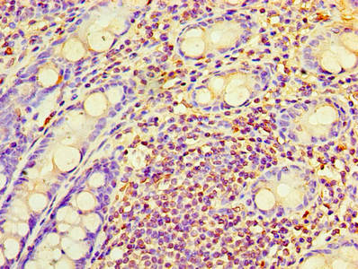
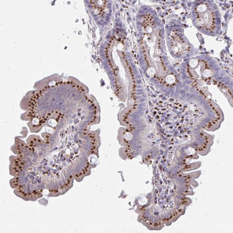
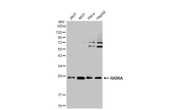
TargetRAB6A
Overview
- SupplierAdipoGen Life Sciences
- Product Nameanti-Rab6-GTP, mAb (rec.) (AA2)
- Delivery Days Customer10
- ApplicationsImmunoCytoChemistry
- CertificationResearch Use Only
- ClonalityMonoclonal
- Clone IDAA2
- Concentration1 mg/ml
- Estimated Purity>95%
- Gene ID5870
- Target nameRAB6A
- Target descriptionRAB6A, member RAS oncogene family
- Target synonymsRAB6, ras-related protein Rab-6A, RAB6, member RAS oncogene family, Rab GTPase
- HostHuman
- IsotypeIgG2
- Protein IDP20340
- Protein NameRas-related protein Rab-6A
- Scientific DescriptionRab6 is involved in protein transport. It is a regulator of membrane traffic from the Golgi apparatus towards the endoplasmic reticulum (ER). - Recombinant Antibody. Recognizes human, mouse and Drosophila GTP-bound Rab6a and Rab6b and mutant Rab6Q72L. Does not detect Rab6 GDP. Species cross-reactivity: Drosophila, Human, Mouse. Clone: AA2. Isotype: Human IgG2lambda. Applications: ICC. Host: Purified from HEK 293 cell culture supernatant. Liquid. In PBS containing 10% glycerol and 0.02% sodium azide. Rab6 is involved in protein transport. It is a regulator of membrane traffic from the Golgi apparatus towards the endoplasmic reticulum (ER).
- ReactivityDrosophila, Human, Mouse
- Storage Instruction-20°C,2°C to 8°C
- UNSPSC41116161
Related products
Product group Antibodies
Anti-RAB6A AntibodyA97306
ApplicationsWestern Blot, ELISA
ReactivityHuman, Mouse, Rat
- SizePrice
Product group Antibodies
anti-Rab6-GTP, mAb (rec.) (AA2) (ATTO 488)AG-27B-0004TD
ApplicationsImmunoCytoChemistry
ReactivityDrosophila, Human, Mouse
TargetRAB6A
- SizePrice
Product group Antibodies
Anti-RAB6A Antibody Picoband(r)A02911-1-CARRIER-FREE
ApplicationsWestern Blot, ImmunoHistoChemistry
ReactivityHuman, Mouse, Rat
TargetRAB6A
- SizePrice
Product group Antibodies
RAB6A Polyclonal AntibodyBS-24347R
ApplicationsWestern Blot
ReactivityBovine, Canine, Equine, Human, Mouse, Porcine, Rabbit, Rat, Sheep
TargetRAB6A
- SizePrice
Product group Antibodies
RAB6A AntibodyCSB-PA019216HA01HU
ApplicationsELISA, ImmunoHistoChemistry
ReactivityHuman
TargetRAB6A
- SizePrice
Product group Antibodies

RAB6A / RAB6 AntibodyLS-C402133
ApplicationsWestern Blot, ELISA, ImmunoHistoChemistry
ReactivityHuman, Mouse, Rat
TargetRAB6A
- SizePrice
Product group Antibodies
Anti-RAB6A-25ulHPA059131
ApplicationsWestern Blot, ImmunoCytoChemistry, ImmunoHistoChemistry
ReactivityHuman
- SizePrice
Product group Antibodies
RAB6A antibodyGTX110646
ApplicationsImmunoPrecipitation, Western Blot, ImmunoHistoChemistry, ImmunoHistoChemistry Paraffin
ReactivityHuman, Mouse, Rat
TargetRAB6A
- SizePrice