


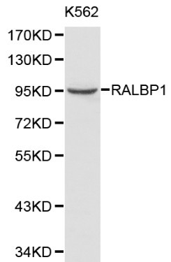
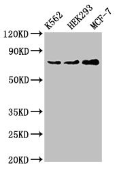
Figure 1. Western blot analysis of RALBP1 using anti-RALBP1 antibody (M01403-2). Electrophoresis was performed on a 5-20% SDS-PAGE gel at 70V (Stacking gel) / 90V (Resolving gel) for 2-3 hours. The sample well of each lane was loaded with 30ug of sample under reducing conditions. Lane 1: human HL-60 whole cell lysates, Lane 2: human U20S whole cell lysates, Lane 3: human Daudi whole cell lysates, Lane 4: human A431 whole cell lysates. After Electrophoresis, proteins were transferred to a Nitrocellulose membrane at 150mA for 50-90 minutes. Blocked the membrane with 5% Non-fat Milk/ TBS for 1.5 hour at RT. The membrane was incubated with mouse anti-RALBP1 antigen affinity purified monoclonal antibody (Catalog # M01403-2) at 0.5 microg/mL overnight at 4°C, then washed with TBS-0.1%Tween 3 times with 5 minutes each and probed with a goat anti-mouse IgG-HRP secondary antibody at a dilution of 1:10000 for 1.5 hour at RT. The signal is developed using an Enhanced Chemiluminescent detection (ECL) kit (Catalog # EK1001) with Tanon 5200 system. A specific band was detected for RALBP1 at approximately 95KD. The expected band size for RALBP1 is at 95KD.
. Overlay histogram showing U87 cells stained with M01403-2 (Blue line). To facilitate intracellular staining, cells were fixed with 4% paraformaldehyde and permeabilized with permeabilization buffer. The cells were blocked with 10% normal goat serum. And then incubated with mouse anti-RALBP1 Antibody (M01403-2, 1microg/1x106 cells) for 30 min at 20°C. DyLight®488 conjugated goat anti-mouse IgG (BA1126, 5-10microg/1x106 cells) was used as secondary antibody for 30 minutes at 20°C. Isotype control antibody (Green line) was mouse IgG (1microg/1x106) used under the same conditions. Unlabelled sample without incubation with primary antibody and secondary antibody (Red line) was used as a blank control.")
Figure 1. Western blot analysis of RALBP1 using anti-RALBP1 antibody (M01403-2). Electrophoresis was performed on a 5-20% SDS-PAGE gel at 70V (Stacking gel) / 90V (Resolving gel) for 2-3 hours. The sample well of each lane was loaded with 30ug of sample under reducing conditions. Lane 1: human HL-60 whole cell lysates, Lane 2: human U20S whole cell lysates, Lane 3: human Daudi whole cell lysates, Lane 4: human A431 whole cell lysates. After Electrophoresis, proteins were transferred to a Nitrocellulose membrane at 150mA for 50-90 minutes. Blocked the membrane with 5% Non-fat Milk/ TBS for 1.5 hour at RT. The membrane was incubated with mouse anti-RALBP1 antigen affinity purified monoclonal antibody (Catalog # M01403-2) at 0.5 microg/mL overnight at 4°C, then washed with TBS-0.1%Tween 3 times with 5 minutes each and probed with a goat anti-mouse IgG-HRP secondary antibody at a dilution of 1:10000 for 1.5 hour at RT. The signal is developed using an Enhanced Chemiluminescent detection (ECL) kit (Catalog # EK1001) with Tanon 5200 system. A specific band was detected for RALBP1 at approximately 95KD. The expected band size for RALBP1 is at 95KD.
Anti-RALBP1 Antibody Picoband(r) (monoclonal, 2E11)
M01403-2-DYLIGHT594
ApplicationsFlow Cytometry, Western Blot
Product group Antibodies
TargetRALBP1
Overview
- SupplierBoster Bio
- Product NameAnti-RALBP1 Antibody Picoband(r) (monoclonal, 2E11)
- Delivery Days Customer9
- ApplicationsFlow Cytometry, Western Blot
- CertificationResearch Use Only
- ClonalityMonoclonal
- Clone ID2E11
- Concentration500 ug/ml
- ConjugateOther Conjugate
- Gene ID10928
- Target nameRALBP1
- Target descriptionralA binding protein 1
- Target synonyms76 kDa Ral-interacting protein; dinitrophenyl S-glutathione ATPase; DNP-SG ATPase; ralA-binding protein 1; ral-interacting protein 1; RIP1; RLIP1; RLIP76
- HostMouse
- IsotypeIgG2a
- Protein IDQ15311
- Protein NameRalA-binding protein 1
- Scientific DescriptionBoster Bio Anti-RALBP1 Antibody Picoband® (monoclonal, 2E11) catalog # M01403-2. Tested in Flow Cytometry, WB applications. This antibody reacts with Human. The brand Picoband indicates this is a premium antibody that guarantees superior quality, high affinity, and strong signals with minimal background in Western blot applications. Only our best-performing antibodies are designated as Picoband, ensuring unmatched performance.
- Storage Instruction-20°C,2°C to 8°C
- UNSPSC12352203
Related products
Product group Antibodies
ApplicationsImmunoPrecipitation
TargetRALBP1
- SizePrice

Product group Antibodies
Anti-RALBP1 Antibody Picoband(r)A01403-1-CARRIER-FREE
ApplicationsFlow Cytometry, ImmunoFluorescence, Western Blot, ELISA, ImmunoCytoChemistry, ImmunoHistoChemistry
TargetRALBP1
- SizePrice
Product group Antibodies
RalBP1 antibodyGTX55774
ApplicationsWestern Blot, ImmunoHistoChemistry, ImmunoHistoChemistry Paraffin
TargetRALBP1
- SizePrice
Product group Antibodies
RALBP1 Polyclonal AntibodyCAC14895
ApplicationsImmunoFluorescence, Western Blot, ELISA, ImmunoHistoChemistry
TargetRALBP1
- SizePrice
Product group Antibodies
Anti-RALBP1 AntibodyA29421
ApplicationsWestern Blot, ImmunoHistoChemistry
- SizePrice
Product group Antibodies
RALBP1 Recombinant AntibodyBSM-61265R
ApplicationsFlow Cytometry, ImmunoFluorescence, Western Blot, ImmunoHistoChemistry, ImmunoHistoChemistry Frozen, ImmunoHistoChemistry Paraffin
TargetRALBP1
- SizePrice
Product group Antibodies
RALBP1 AntibodyCSB-PA618981LA01HU
ApplicationsImmunoFluorescence, Western Blot, ELISA, ImmunoHistoChemistry
ReactivityHuman
TargetRALBP1
- SizePrice

Product group Antibodies
Anti-RALBP1 AntibodyHPA007622
ApplicationsImmunoCytoChemistry
ReactivityHuman
TargetRALBP1
- SizePrice