


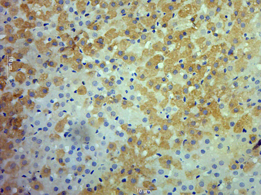
Immunohistochemical staining of human adrenal gland shows strong membranous positivity in glandular cells.

Immunohistochemical staining of human adrenal gland shows strong membranous positivity in glandular cells.
Anti-RET Antibody
HPA008356
ApplicationsImmunoCytoChemistry, ImmunoHistoChemistry
Product group Antibodies
ReactivityHuman
TargetRET
Overview
- SupplierAtlas Antibodies
- Product NameAnti-RET Antibody
- Delivery Days Customer4
- ApplicationsImmunoCytoChemistry, ImmunoHistoChemistry
- CertificationResearch Use Only
- ClonalityPolyclonal
- ConjugateUnconjugated
- Gene ID5979
- Target nameRET
- Target descriptionret proto-oncogene
- Target synonymsCDHF12, CDHR16, HSCR1, MEN2A, MEN2B, MTC1, PTC, RET-ELE1, proto-oncogene tyrosine-protein kinase receptor Ret, RET receptor tyrosine kinase, cadherin family member 12, cadherin-related family member 16, proto-oncogene c-Ret, rearranged during transfection, ret proto-oncogene (multiple endocrine neoplasia and medullary thyroid carcinoma 1, Hirschsprung disease)
- HostRabbit
- IsotypeIgG
- Protein IDP07949
- Protein NameProto-oncogene tyrosine-protein kinase receptor Ret
- Scientific DescriptionRecombinant Protein Epitope Signature Tag (PrEST) antigen sequence
- ReactivityHuman
- Storage Instruction-20°C,2°C to 8°C
- UNSPSC41116161
Datasheet
MSDS
Related products
Product group Antibodies
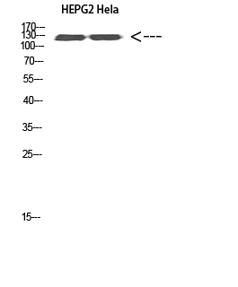
Anti-RET AntibodyA100044
ApplicationsWestern Blot, ELISA, ImmunoHistoChemistry
ReactivityHuman
- SizePrice
Product group Antibodies

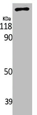
RET AntibodyLS-C761146
ApplicationsWestern Blot
ReactivityHuman, Mouse, Rat
TargetRET
- SizePrice

Product group Antibodies
Anti-RET Antibody Picoband(r)A00293-1-CARRIER-FREE
ApplicationsWestern Blot, ELISA, ImmunoHistoChemistry
ReactivityHuman, Mouse, Rat
TargetRET
- SizePrice
Product group Antibodies
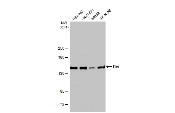
RET Polyclonal AntibodyBS-2793R
ApplicationsImmunoFluorescence, Western Blot, ELISA, ImmunoCytoChemistry, ImmunoHistoChemistry, ImmunoHistoChemistry Frozen, ImmunoHistoChemistry Paraffin
ReactivityCanine, Human, Mouse, Rabbit, Rat
TargetRET
- SizePrice
Product group Antibodies
RET AntibodyCSB-PA003961
ApplicationsImmunoFluorescence, Western Blot, ELISA, ImmunoHistoChemistry
ReactivityHuman, Mouse, Rat
TargetRET
- SizePrice
Product group Antibodies
Ret Recombinant AntibodyCAC12550
ApplicationsFlow Cytometry, ELISA
TargetRET
- SizePrice
Product group Antibodies
Ret antibodyGTX101059
ApplicationsWestern Blot
ReactivityHuman
TargetRET
- SizePrice