


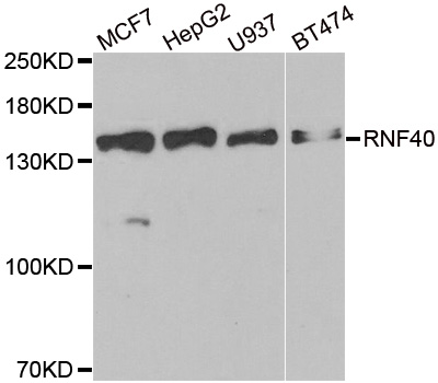
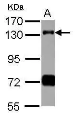
Figure 1. Western blot analysis of RNF40 using anti-RNF40 antibody (A06979-2). Electrophoresis was performed on a 5-20% SDS-PAGE gel at 70V (Stacking gel) / 90V (Resolving gel) for 2-3 hours. The sample well of each lane was loaded with 30 ug of sample under reducing conditions. Lane 1: human 293T whole cell lysates, Lane 2: human Hela whole cell lysates, Lane 3: human MCF-7 whole cell lysates. After electrophoresis, proteins were transferred to a nitrocellulose membrane at 150 mA for 50-90 minutes. Blocked the membrane with 5% non-fat milk/TBS for 1.5 hour at RT. The membrane was incubated with rabbit anti-RNF40 antigen affinity purified polyclonal antibody (Catalog # A06979-2) at 0.5 microg/mL overnight at 4°C, then washed with TBS-0.1%Tween 3 times with 5 minutes each and probed with a goat anti-rabbit IgG-HRP secondary antibody at a dilution of 1:5000 for 1.5 hour at RT. The signal is developed using an Enhanced Chemiluminescent detection (ECL) kit (Catalog # EK1002) with Tanon 5200 system. A specific band was detected for RNF40 at approximately 130 kDa. The expected band size for RNF40 is at 114 kDa.
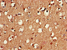
. RNF40 was detected in a paraffin-embedded section of human liver cancer tissue. Heat mediated antigen retrieval was performed in EDTA buffer (pH 8.0, epitope retrieval solution). The tissue section was blocked with 10% goat serum. The tissue section was then incubated with 2 microg/ml rabbit anti-RNF40 Antibody (A06979-2) overnight at 4°C. Peroxidase Conjugated Goat Anti-rabbit IgG was used as secondary antibody and incubated for 30 minutes at 37°C. The tissue section was developed using HRP Conjugated Rabbit IgG Super Vision Assay Kit (Catalog # SV0002) with DAB as the chromogen.")
. RNF40 was detected in a paraffin-embedded section of human lung adenocarcinoma tissue. Heat mediated antigen retrieval was performed in EDTA buffer (pH 8.0, epitope retrieval solution). The tissue section was blocked with 10% goat serum. The tissue section was then incubated with 2 microg/ml rabbit anti-RNF40 Antibody (A06979-2) overnight at 4°C. Peroxidase Conjugated Goat Anti-rabbit IgG was used as secondary antibody and incubated for 30 minutes at 37°C. The tissue section was developed using HRP Conjugated Rabbit IgG Super Vision Assay Kit (Catalog # SV0002) with DAB as the chromogen.")
. RNF40 was detected in a paraffin-embedded section of human testicular seminoma tissue. Heat mediated antigen retrieval was performed in EDTA buffer (pH 8.0, epitope retrieval solution). The tissue section was blocked with 10% goat serum. The tissue section was then incubated with 2 microg/ml rabbit anti-RNF40 Antibody (A06979-2) overnight at 4°C. Peroxidase Conjugated Goat Anti-rabbit IgG was used as secondary antibody and incubated for 30 minutes at 37°C. The tissue section was developed using HRP Conjugated Rabbit IgG Super Vision Assay Kit (Catalog # SV0002) with DAB as the chromogen.")
. RNF40 was detected in a paraffin-embedded section of human lung cancer tissue. Heat mediated antigen retrieval was performed in EDTA buffer (pH 8.0, epitope retrieval solution). The tissue section was blocked with 10% goat serum. The tissue section was then incubated with 5 microg/mL rabbit anti-RNF40 Antibody (A06979-2) overnight at 4°C. DyLight®550 Conjugated Goat Anti-Rabbit IgG (BA1135) was used as secondary antibody at 1:500 dilution and incubated for 30 minutes at 37°C. Visualize using a fluorescence microscope and filter sets appropriate for the label used.")
Figure 1. Western blot analysis of RNF40 using anti-RNF40 antibody (A06979-2). Electrophoresis was performed on a 5-20% SDS-PAGE gel at 70V (Stacking gel) / 90V (Resolving gel) for 2-3 hours. The sample well of each lane was loaded with 30 ug of sample under reducing conditions. Lane 1: human 293T whole cell lysates, Lane 2: human Hela whole cell lysates, Lane 3: human MCF-7 whole cell lysates. After electrophoresis, proteins were transferred to a nitrocellulose membrane at 150 mA for 50-90 minutes. Blocked the membrane with 5% non-fat milk/TBS for 1.5 hour at RT. The membrane was incubated with rabbit anti-RNF40 antigen affinity purified polyclonal antibody (Catalog # A06979-2) at 0.5 microg/mL overnight at 4°C, then washed with TBS-0.1%Tween 3 times with 5 minutes each and probed with a goat anti-rabbit IgG-HRP secondary antibody at a dilution of 1:5000 for 1.5 hour at RT. The signal is developed using an Enhanced Chemiluminescent detection (ECL) kit (Catalog # EK1002) with Tanon 5200 system. A specific band was detected for RNF40 at approximately 130 kDa. The expected band size for RNF40 is at 114 kDa.
Anti-RNF40 Antibody Picoband(r)
A06979-2-CARRIER-FREE
ApplicationsImmunoFluorescence, Western Blot, ELISA, ImmunoHistoChemistry
Product group Antibodies
ReactivityHuman
TargetRNF40
Overview
- SupplierBoster Bio
- Product NameAnti-RNF40 Antibody Picoband(r)
- Delivery Days Customer9
- ApplicationsImmunoFluorescence, Western Blot, ELISA, ImmunoHistoChemistry
- CertificationResearch Use Only
- ClonalityPolyclonal
- Concentration500 ug/ml
- Gene ID9810
- Target nameRNF40
- Target descriptionring finger protein 40
- Target synonymsBRE1B, RBP95, STARING, E3 ubiquitin-protein ligase BRE1B, 95 kDa retinoblastoma protein binding protein, 95 kDa retinoblastoma-associated protein, BRE1 E3 ubiquitin ligase homolog B, BRE1-B, RING-type E3 ubiquitin transferase BRE1B, Rb-associated protein, ring finger protein 40, E3 ubiquitin protein ligase
- HostRabbit
- IsotypeIgG
- Protein IDO75150
- Protein NameE3 ubiquitin-protein ligase BRE1B
- Scientific DescriptionBoster Bio Anti-RNF40 Antibody Picoband® catalog # A06979-2. Tested in ELISA, IF, IHC, WB applications. This antibody reacts with Human. The brand Picoband indicates this is a premium antibody that guarantees superior quality, high affinity, and strong signals with minimal background in Western blot applications. Only our best-performing antibodies are designated as Picoband, ensuring unmatched performance.
- ReactivityHuman
- Storage Instruction-20°C,2°C to 8°C
- UNSPSC12352203
Related products
Product group Antibodies
Anti-RNF40 AntibodyA31387
ApplicationsWestern Blot, ImmunoHistoChemistry
ReactivityHuman, Mouse, Rat
- SizePrice
Product group Antibodies
Anti-RNF40 Antibody144-06443
ApplicationsImmunoFluorescence, Western Blot, ChIP Chromatin ImmunoPrecipitation, ImmunoHistoChemistry
ReactivityHuman
TargetRNF40
- SizePrice
Product group Antibodies
RNF40 Recombinant AntibodyBSM-62002R
ApplicationsImmunoFluorescence, ImmunoPrecipitation, Western Blot, ImmunoHistoChemistry, ImmunoHistoChemistry Frozen, ImmunoHistoChemistry Paraffin
ReactivityHuman, Mouse, Rat
TargetRNF40
- SizePrice
Product group Antibodies
RNF40 AntibodyCSB-PA019890LA01HU
ApplicationsELISA, ImmunoHistoChemistry
ReactivityHuman
TargetRNF40
- SizePrice
Product group Antibodies

RNF40 / STARING AntibodyLS-C334722
ApplicationsImmunoFluorescence, Western Blot, ImmunoHistoChemistry
ReactivityHuman
TargetRNF40
- SizePrice

Product group Antibodies
Anti-RNF40 AntibodyHPA041330
ApplicationsWestern Blot, ImmunoHistoChemistry
ReactivityHuman, Mouse, Rat
TargetRNF40
- SizePrice
Product group Antibodies
RNF40 antibody [N3C1-2], InternalGTX115294
ApplicationsWestern Blot
ReactivityHuman
TargetRNF40
- SizePrice
Product group Antibodies
Anti-RNF40 AntibodyCAB6443
ApplicationsWestern Blot, ELISA
ReactivityHuman
TargetRNF40
- SizePrice