


Figure 1. Western blot analysis of RRM2 using anti-RRM2 antibody (PB9817). Electrophoresis was performed on a 5-20% SDS-PAGE gel at 70V (Stacking gel) / 90V (Resolving gel) for 2-3 hours. Lane 1: Rat Cardiac Muscle Tissue Lysate at 50ug, Lane 2: Mouse Cardiac Muscle Tissue Lysate at 50ug, Lane 3: A431 Whole Cell Lysate at 40ug, Lane 4: HELA Whole Cell Lysate at 40ug. After electrophoresis, proteins were transferred to a nitrocellulose membrane at 150 mA for 50-90 minutes. Blocked the membrane with 5% non-fat milk/TBS for 1.5 hour at RT. The membrane was incubated with rabbit anti-RRM2 antigen affinity purified polyclonal antibody (Catalog # PB9817) at 0.5 microg/mL overnight at 4°C, then washed with TBS-0.1%Tween 3 times with 5 minutes each and probed with a goat anti-rabbit IgG-HRP secondary antibody at a dilution of 1:5000 for 1.5 hour at RT. The signal is developed using an Enhanced Chemiluminescent detection (ECL) kit (Catalog # EK1002) with Tanon 5200 system. A specific band was detected for RRM2 at approximately 50 kDa. The expected band size for RRM2 is at 50 kDa.
. RRM2 was detected in a paraffin-embedded section of human mammary cancer tissue. Heat mediated antigen retrieval was performed in EDTA buffer (pH 8.0, epitope retrieval solution). The tissue section was blocked with 10% goat serum. The tissue section was then incubated with 1 microg/ml rabbit anti-RRM2 Antibody (PB9817) overnight at 4°C. Biotinylated goat anti-rabbit IgG was used as secondary antibody and incubated for 30 minutes at 37°C. The tissue section was developed using Strepavidin-Biotin-Complex (SABC) (Catalog # SA1022) with DAB as the chromogen.")
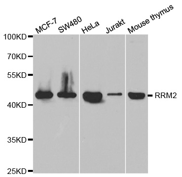
Figure 1. Western blot analysis of RRM2 using anti-RRM2 antibody (PB9817). Electrophoresis was performed on a 5-20% SDS-PAGE gel at 70V (Stacking gel) / 90V (Resolving gel) for 2-3 hours. Lane 1: Rat Cardiac Muscle Tissue Lysate at 50ug, Lane 2: Mouse Cardiac Muscle Tissue Lysate at 50ug, Lane 3: A431 Whole Cell Lysate at 40ug, Lane 4: HELA Whole Cell Lysate at 40ug. After electrophoresis, proteins were transferred to a nitrocellulose membrane at 150 mA for 50-90 minutes. Blocked the membrane with 5% non-fat milk/TBS for 1.5 hour at RT. The membrane was incubated with rabbit anti-RRM2 antigen affinity purified polyclonal antibody (Catalog # PB9817) at 0.5 microg/mL overnight at 4°C, then washed with TBS-0.1%Tween 3 times with 5 minutes each and probed with a goat anti-rabbit IgG-HRP secondary antibody at a dilution of 1:5000 for 1.5 hour at RT. The signal is developed using an Enhanced Chemiluminescent detection (ECL) kit (Catalog # EK1002) with Tanon 5200 system. A specific band was detected for RRM2 at approximately 50 kDa. The expected band size for RRM2 is at 50 kDa.
Anti-RRM2 Antibody Picoband(r)
PB9817-IFLUOR647
ApplicationsWestern Blot, ImmunoHistoChemistry
Product group Antibodies
ReactivityHuman, Mouse, Rat
TargetRRM2
Overview
- SupplierBoster Bio
- Product NameAnti-RRM2 Antibody Picoband(r)
- Delivery Days Customer9
- Application Supplier NoteTested Species: In-house tested species with positive results. By Heat: Boiling the paraffin sections in 10mM citrate buffer, pH6.0, for 20mins is required for the staining of formalin/paraffin sections. Other applications have not been tested. Optimal dilutions should be determined by end users.
- ApplicationsWestern Blot, ImmunoHistoChemistry
- CertificationResearch Use Only
- ClonalityPolyclonal
- Concentration500 ug/ml
- ConjugateOther Conjugate
- Gene ID6241
- Target nameRRM2
- Target descriptionribonucleotide reductase regulatory subunit M2
- Target synonymsC2orf48, R2, RR2, RR2M, ribonucleoside-diphosphate reductase subunit M2, ribonucleotide reductase M2 polypeptide, ribonucleotide reductase small chain, ribonucleotide reductase small subunit, uncharacterized protein C2orf48
- HostRabbit
- IsotypeIgG
- Protein IDP31350
- Protein NameRibonucleoside-diphosphate reductase subunit M2
- Scientific DescriptionBoster Bio Anti-RRM2 Antibody Picoband® catalog # PB9817. Tested in IHC, WB applications. This antibody reacts with Human, Mouse, Rat. The brand Picoband indicates this is a premium antibody that guarantees superior quality, high affinity, and strong signals with minimal background in Western blot applications. Only our best-performing antibodies are designated as Picoband, ensuring unmatched performance.
- ReactivityHuman, Mouse, Rat
- Storage Instruction-20°C,2°C to 8°C
- UNSPSC12352203
Related products
Product group Antibodies
RRM2 Recombinant Antibody, AbBy Fluor-488 ConjugatedBSM-61703R-BF488
ApplicationsImmunoFluorescence, Western Blot, ImmunoCytoChemistry
ReactivityHuman
TargetRRM2
- SizePrice
Product group Antibodies
RRM2 Polyclonal AntibodyCAC13191
ApplicationsWestern Blot, ELISA
TargetRRM2
- SizePrice
Product group Antibodies
Anti-RRM2 AntibodyA30719
ApplicationsWestern Blot, ImmunoHistoChemistry
ReactivityHuman, Mouse, Rat
- SizePrice
Product group Antibodies
Anti-RRM2 Antibody144-05255
ApplicationsImmunoFluorescence, ImmunoPrecipitation, Western Blot, ImmunoHistoChemistry
ReactivityHuman, Mouse, Rat
TargetRRM2
- SizePrice
Product group Antibodies

Anti-RRM2 [SAIC-30C-18]AB00322-1.1-BT
ApplicationsMass Spectrometry, Western Blot, ELISA
ReactivityHuman
TargetRRM2
- SizePrice
Product group Antibodies
References
RRM2 antibody [N1C1]GTX103193
ApplicationsImmunoFluorescence, Western Blot, ImmunoCytoChemistry, ImmunoHistoChemistry, ImmunoHistoChemistry Paraffin
ReactivityHuman, Mouse, Rat, Zebra Fish
TargetRRM2
- SizePrice
Product group Antibodies

RRM2 AntibodyLS-C333948
ApplicationsImmunoFluorescence, ImmunoPrecipitation, Western Blot, ImmunoHistoChemistry
ReactivityHuman, Mouse, Rat
TargetRRM2
- SizePrice

Product group Antibodies
Anti-RRM2 AntibodyHPA056994
ApplicationsWestern Blot, ImmunoCytoChemistry, ImmunoHistoChemistry
ReactivityHuman
TargetRRM2
- SizePrice