

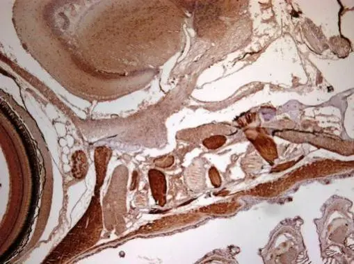
Immunohistochemical analysis of paraffin-embedded zebrafish tissue, using RRM2 antibody [N1C1] (GTX103193) at 1:300 dilution.
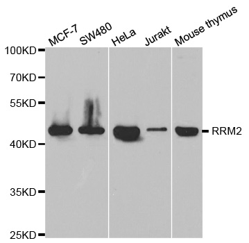
![Various tissue extracts (30 μg) were separated by 10% SDS-PAGE, and the membrane was blotted with RRM2 antibody [N1C1] (GTX103193) diluted at 1:1000.](https://www.genetex.com/upload/website/prouct_img/normal/GTX103193/GTX103193_42718_20170105_WB_Z_22111423_302.webp "Various tissue extracts (30 μg) were separated by 10% SDS-PAGE, and the membrane was blotted with RRM2 antibody [N1C1] (GTX103193) diluted at 1:1000.")
![RRM2 antibody [N1C1] detects RRM2 protein at cytoplasm in rat intestine by immunohistochemical analysis. Sample: Paraffin-embedded rat intestine. RRM2 antibody [N1C1] (GTX103193) diluted at 1:500.
Antigen Retrieval: Citrate buffer, pH 6.0, 15 min](https://www.genetex.com/upload/website/prouct_img/normal/GTX103193/GTX103193_42151_20170322_IHC-P_R_w_23060119_791.webp "RRM2 antibody [N1C1] detects RRM2 protein at cytoplasm in rat intestine by immunohistochemical analysis. Sample: Paraffin-embedded rat intestine. RRM2 antibody [N1C1] (GTX103193) diluted at 1:500.
Antigen Retrieval: Citrate buffer, pH 6.0, 15 min")
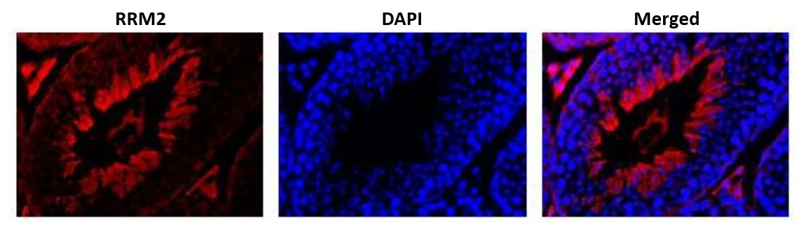
![RRM2 antibody [N1C1] detects RRM2 protein at cytoplasm by immunofluorescent analysis. Sample: HeLa cells were fixed in ice-cold MeOH for 5 min. Green: RRM2 protein stained by RRM2 antibody [N1C1] (GTX103193) diluted at 1:1000. Blue: Hoechst 33342 staining. Scale bar = 10 μm.](https://www.genetex.com/upload/website/prouct_img/normal/GTX103193/GTX103193_42718_20161123_IFA_w_23060119_243.webp "RRM2 antibody [N1C1] detects RRM2 protein at cytoplasm by immunofluorescent analysis. Sample: HeLa cells were fixed in ice-cold MeOH for 5 min. Green: RRM2 protein stained by RRM2 antibody [N1C1] (GTX103193) diluted at 1:1000. Blue: Hoechst 33342 staining. Scale bar = 10 μm.")
![RRM2 antibody [N1C1] detects RRM2 protein at cytoplasm on mouse muscle by immunohistochemical analysis. Sample: Paraffin-embedded mouse muscle. RRM2 antibody [N1C1] (GTX103193) diluted at 1:500.
Antigen Retrieval: Trilogy? (EDTA based, pH 8.0) buffer, 15min](https://www.genetex.com/upload/website/prouct_img/normal/GTX103193/GTX103193_40128_20141108_IHC_M_2_w_23060119_739.webp "RRM2 antibody [N1C1] detects RRM2 protein at cytoplasm on mouse muscle by immunohistochemical analysis. Sample: Paraffin-embedded mouse muscle. RRM2 antibody [N1C1] (GTX103193) diluted at 1:500.
Antigen Retrieval: Trilogy? (EDTA based, pH 8.0) buffer, 15min")
![RRM2 antibody [N1C1] detects RRM2 protein at cytoplasm by immunofluorescent analysis. Sample: HeLa cells were fixed in 4% paraformaldehyde at RT for 15 min. Green: RRM2 protein stained by RRM2 antibody [N1C1] (GTX103193) diluted at 1:500. Blue: Hoechst 33342 staining. Scale bar = 10 μm.](https://www.genetex.com/upload/website/prouct_img/normal/GTX103193/GTX103193_41745_20150513_IFA_w_23060119_428.webp "RRM2 antibody [N1C1] detects RRM2 protein at cytoplasm by immunofluorescent analysis. Sample: HeLa cells were fixed in 4% paraformaldehyde at RT for 15 min. Green: RRM2 protein stained by RRM2 antibody [N1C1] (GTX103193) diluted at 1:500. Blue: Hoechst 33342 staining. Scale bar = 10 μm.")
![RRM2 antibody [N1C1] detects RRM2 protein at cytoplasm in mouse prostate by immunohistochemical analysis. Sample: Paraffin-embedded mouse muscle. RRM2 antibody [N1C1] (GTX103193) diluted at 1:500.
Antigen Retrieval: Citrate buffer, pH 6.0, 15 min](https://www.genetex.com/upload/website/prouct_img/normal/GTX103193/GTX103193_42718_20170322_IHC-P_M_w_23060119_725.webp "RRM2 antibody [N1C1] detects RRM2 protein at cytoplasm in mouse prostate by immunohistochemical analysis. Sample: Paraffin-embedded mouse muscle. RRM2 antibody [N1C1] (GTX103193) diluted at 1:500.
Antigen Retrieval: Citrate buffer, pH 6.0, 15 min")
![RRM2 antibody [N1C1] detects RRM2 protein at cytoplasm in rat intestine by immunohistochemical analysis. Sample: Paraffin-embedded rat intestine. RRM2 antibody [N1C1] (GTX103193) diluted at 1:500.
Antigen Retrieval: Citrate buffer, pH 6.0, 15 min](https://www.genetex.com/upload/website/prouct_img/normal/GTX103193/GTX103193_42718_20170322_IHC-P_R_w_23060119_638.webp "RRM2 antibody [N1C1] detects RRM2 protein at cytoplasm in rat intestine by immunohistochemical analysis. Sample: Paraffin-embedded rat intestine. RRM2 antibody [N1C1] (GTX103193) diluted at 1:500.
Antigen Retrieval: Citrate buffer, pH 6.0, 15 min")
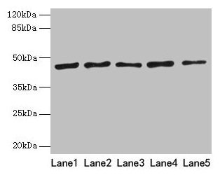
![Various whole cell extracts (30 μg) were separated by 10% SDS-PAGE, and the membrane was blotted with RRM2 antibody [N1C1] (GTX103193) diluted at 1:1000. The HRP-conjugated anti-rabbit IgG antibody (GTX213110-01) was used to detect the primary antibody. Corresponding RNA expression data for the same cell lines are based on Human Protein Atlas program.](https://www.genetex.com/upload/website/prouct_img/normal/GTX103193/GTX103193_44265_20210402_WB_TPM_watermark_23121122_855.webp "Various whole cell extracts (30 μg) were separated by 10% SDS-PAGE, and the membrane was blotted with RRM2 antibody [N1C1] (GTX103193) diluted at 1:1000. The HRP-conjugated anti-rabbit IgG antibody (GTX213110-01) was used to detect the primary antibody. Corresponding RNA expression data for the same cell lines are based on Human Protein Atlas program.")

Immunohistochemical analysis of paraffin-embedded zebrafish tissue, using RRM2 antibody [N1C1] (GTX103193) at 1:300 dilution.
RRM2 antibody [N1C1]
GTX103193
ApplicationsImmunoFluorescence, Western Blot, ImmunoCytoChemistry, ImmunoHistoChemistry, ImmunoHistoChemistry Paraffin
Product group Antibodies
ReactivityHuman, Mouse, Rat, Zebra Fish
TargetRRM2
Overview
- SupplierGeneTex
- Product NameRRM2 antibody [N1C1]
- Delivery Days Customer9
- Application Supplier NoteICC/IF: 1:100-1:1000. IHC-P: 1:100-1:1000. *Optimal dilutions/concentrations should be determined by the researcher.Not tested in other applications.
- ApplicationsImmunoFluorescence, Western Blot, ImmunoCytoChemistry, ImmunoHistoChemistry, ImmunoHistoChemistry Paraffin
- CertificationResearch Use Only
- ClonalityPolyclonal
- Concentration0.28 mg/ml
- ConjugateUnconjugated
- Gene ID6241
- Target nameRRM2
- Target descriptionribonucleotide reductase regulatory subunit M2
- Target synonymsC2orf48, R2, RR2, RR2M, ribonucleoside-diphosphate reductase subunit M2, ribonucleotide reductase M2 polypeptide, ribonucleotide reductase small chain, ribonucleotide reductase small subunit, uncharacterized protein C2orf48
- HostRabbit
- IsotypeIgG
- Protein IDP31350
- Protein NameRibonucleoside-diphosphate reductase subunit M2
- Scientific DescriptionThis gene encodes one of two non-identical subunits for ribonucleotide reductase. This reductase catalyzes the formation of deoxyribonucleotides from ribonucleotides. Synthesis of the encoded protein (M2) is regulated in a cell-cycle dependent fashion. [provided by RefSeq]
- ReactivityHuman, Mouse, Rat, Zebra Fish
- Storage Instruction-20°C or -80°C,2°C to 8°C
- UNSPSC41116161
Datasheet
Related products
Product group Antibodies
Anti-RRM2 AntibodyA30719
ApplicationsWestern Blot, ImmunoHistoChemistry
ReactivityHuman, Mouse, Rat
- SizePrice
Product group Antibodies

Anti-RRM2 [SAIC-30C-18]AB00322-1.1-BT
ApplicationsMass Spectrometry, Western Blot, ELISA
ReactivityHuman
TargetRRM2
- SizePrice
Product group Antibodies
Anti-RRM2 Antibody144-05255
ApplicationsImmunoFluorescence, ImmunoPrecipitation, Western Blot, ImmunoHistoChemistry
ReactivityHuman, Mouse, Rat
TargetRRM2
- SizePrice
Product group Antibodies
RRM2 Recombinant Antibody, AbBy Fluor-488 ConjugatedBSM-61703R-BF488
ApplicationsWestern Blot
ReactivityHuman
TargetRRM2
- SizePrice
Product group Antibodies
RRM2 Polyclonal AntibodyCAC13191
ApplicationsWestern Blot, ELISA
TargetRRM2
- SizePrice
Product group Antibodies
RRM2 AntibodyCSB-PA020519HA01HU
ApplicationsWestern Blot, ELISA
ReactivityHuman
TargetRRM2
- SizePrice
Product group Antibodies
RRM2 antibodyGTX00888
ApplicationsImmunoFluorescence, ImmunoPrecipitation, Western Blot, ImmunoCytoChemistry, ImmunoHistoChemistry, ImmunoHistoChemistry Paraffin
ReactivityHamster, Human, Mouse, Rat, Xenopus
TargetRRM2
- SizePrice
Product group Antibodies

RRM2 AntibodyLS-C333948
ApplicationsImmunoFluorescence, ImmunoPrecipitation, Western Blot, ImmunoHistoChemistry
ReactivityHuman, Mouse, Rat
TargetRRM2
- SizePrice

Product group Antibodies
Anti-RRM2 AntibodyHPA056994
ApplicationsWestern Blot, ImmunoCytoChemistry, ImmunoHistoChemistry
ReactivityHuman
TargetRRM2
- SizePrice
![Whole zebrafish extract (30 μg) was separated by 10% SDS-PAGE, and the membrane was blotted with RRM2 antibody [HL1235] (GTX636634) diluted at 1:1000. The HRP-conjugated anti-rabbit IgG antibody (GTX213110-01) was used to detect the primary antibody.](https://www.genetex.com/upload/website/prouct_img/normal/GTX636634/GTX636634_44564_20221216_WB_Z_22122018_344.webp)
Product group Antibodies
RRM2 antibody [HL1235]GTX636634
ApplicationsImmunoFluorescence, Western Blot, ImmunoCytoChemistry, ImmunoHistoChemistry, ImmunoHistoChemistry Paraffin
ReactivityHuman, Mouse, Zebra Fish
TargetRRM2
- SizePrice