

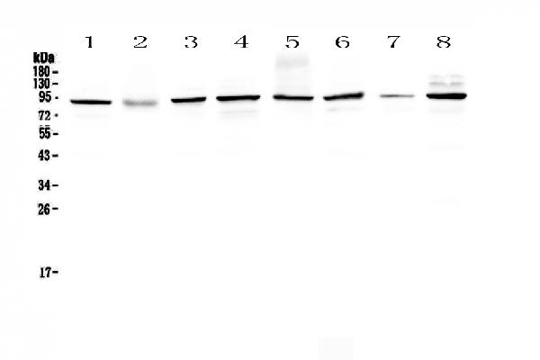
Figure 1. Western blot analysis of SAE2/UBA2 using anti-SAE2/UBA2 antibody (A03816-2). Electrophoresis was performed on a 5-20% SDS-PAGE gel at 70V (Stacking gel) / 90V (Resolving gel) for 2-3 hours. The sample well of each lane was loaded with 50ug of sample under reducing conditions. Lane 1: human Hela whole cell lysates, Lane 2: human placenta tissue lysates, Lane 3: human MCF-7 whole cell lysates, Lane 4: human A549 whole cell lysates, Lane 5: human SK-OV-3 whole cell lysates, Lane 6: human 22RV1 whole cell lysates, Lane 7: human A431 whole cell lysates, Lane 8: human COLO-320 whole cell lysates. After Electrophoresis, proteins were transferred to a Nitrocellulose membrane at 150mA for 50-90 minutes. Blocked the membrane with 5% Non-fat Milk/ TBS for 1.5 hour at RT. The membrane was incubated with rabbit anti-SAE2/UBA2 antigen affinity purified polyclonal antibody (Catalog # A03816-2) at 0.5 microg/mL overnight at 4°C, then washed with TBS-0.1%Tween 3 times with 5 minutes each and probed with a goat anti-rabbit IgG-HRP secondary antibody at a dilution of 1:10000 for 1.5 hour at RT. The signal is developed using an Enhanced Chemiluminescent detection (ECL) kit (Catalog # EK1002) with Tanon 5200 system. A specific band was detected for SAE2/UBA2 at approximately 90KD. The expected band size for SAE2/UBA2 is at 71KD.
. SAE2/UBA2 was detected in paraffin-embedded section of human colon cancer tissue. Heat mediated antigen retrieval was performed in citrate buffer (pH6, epitope retrieval solution) for 20 mins. The tissue section was blocked with 10% goat serum. The tissue section was then incubated with 1microg/ml rabbit anti-SAE2/UBA2 Antibody (A03816-2) overnight at 4°C. Biotinylated goat anti-rabbit IgG was used as secondary antibody and incubated for 30 minutes at 37°C. The tissue section was developed using Strepavidin-Biotin-Complex (SABC)(Catalog # SA1022) with DAB as the chromogen.")
. SAE2/UBA2 was detected in paraffin-embedded section of human mammary cancer tissue. Heat mediated antigen retrieval was performed in citrate buffer (pH6, epitope retrieval solution) for 20 mins. The tissue section was blocked with 10% goat serum. The tissue section was then incubated with 1microg/ml rabbit anti-SAE2/UBA2 Antibody (A03816-2) overnight at 4°C. Biotinylated goat anti-rabbit IgG was used as secondary antibody and incubated for 30 minutes at 37°C. The tissue section was developed using Strepavidin-Biotin-Complex (SABC)(Catalog # SA1022) with DAB as the chromogen.")
. SAE2/UBA2 was detected in paraffin-embedded section of rat testis tissue. Heat mediated antigen retrieval was performed in citrate buffer (pH6, epitope retrieval solution) for 20 mins. The tissue section was blocked with 10% goat serum. The tissue section was then incubated with 1microg/ml rabbit anti-SAE2/UBA2 Antibody (A03816-2) overnight at 4°C. Biotinylated goat anti-rabbit IgG was used as secondary antibody and incubated for 30 minutes at 37°C. The tissue section was developed using Strepavidin-Biotin-Complex (SABC)(Catalog # SA1022) with DAB as the chromogen.")
. SAE2/UBA2 was detected in paraffin-embedded section of mouse testis tissue. Heat mediated antigen retrieval was performed in citrate buffer (pH6, epitope retrieval solution) for 20 mins. The tissue section was blocked with 10% goat serum. The tissue section was then incubated with 1microg/ml rabbit anti-SAE2/UBA2 Antibody (A03816-2) overnight at 4°C. Biotinylated goat anti-rabbit IgG was used as secondary antibody and incubated for 30 minutes at 37°C. The tissue section was developed using Strepavidin-Biotin-Complex (SABC)(Catalog # SA1022) with DAB as the chromogen.")
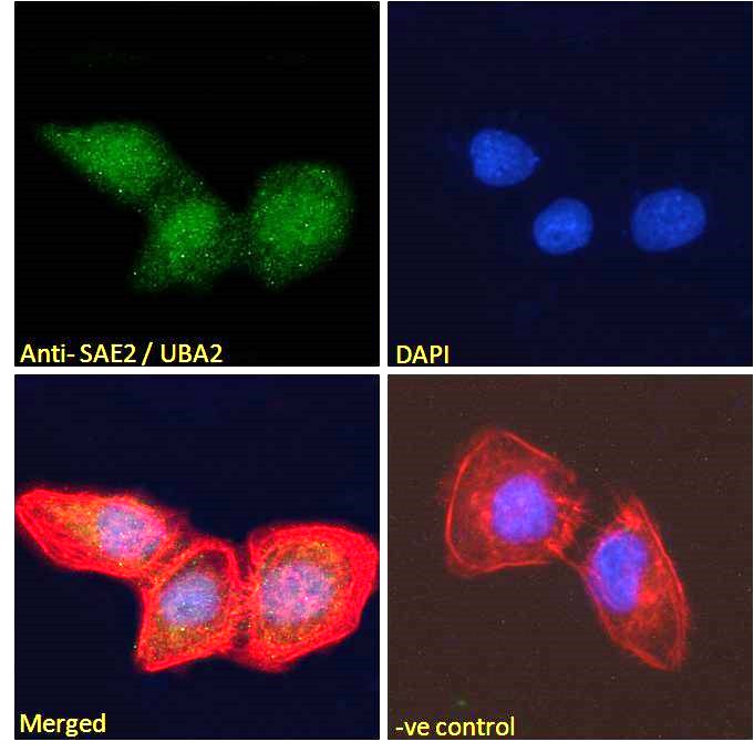
. SAE2/UBA2 was detected in immunocytochemical section of A431 cell. Enzyme antigen retrieval was performed using IHC enzyme antigen retrieval reagent (AR0022) for 15 mins. The cells were blocked with 10% goat serum. And then incubated with 2microg/mL rabbit anti-SAE2/UBA2 Antibody (A03816-2) overnight at 4°C. DyLight®488 Conjugated Goat Anti-Rabbit IgG (BA1127) was used as secondary antibody at 1:100 dilution and incubated for 30 minutes at 37°C. The section was counterstained with DAPI. Visualize using a fluorescence microscope and filter sets appropriate for the label used.")
. Overlay histogram showing A431 cells stained with A03816-2 (Blue line). To facilitate intracellular staining, cells were fixed with 4% paraformaldehyde and permeabilized with permeabilization buffer. The cells were blocked with 10% normal goat serum. And then incubated with rabbit anti-SAE2/UBA2 Antibody (A03816-2,1microg/1x106 cells) for 30 min at 20°C. DyLight®488 conjugated goat anti-rabbit IgG (BA1127, 5-10microg/1x106 cells) was used as secondary antibody for 30 minutes at 20°C. Isotype control antibody (Green line) was rabbit IgG (1microg/1x106) used under the same conditions. Unlabelled sample (Red line) was also used as a control.")
Figure 1. Western blot analysis of SAE2/UBA2 using anti-SAE2/UBA2 antibody (A03816-2). Electrophoresis was performed on a 5-20% SDS-PAGE gel at 70V (Stacking gel) / 90V (Resolving gel) for 2-3 hours. The sample well of each lane was loaded with 50ug of sample under reducing conditions. Lane 1: human Hela whole cell lysates, Lane 2: human placenta tissue lysates, Lane 3: human MCF-7 whole cell lysates, Lane 4: human A549 whole cell lysates, Lane 5: human SK-OV-3 whole cell lysates, Lane 6: human 22RV1 whole cell lysates, Lane 7: human A431 whole cell lysates, Lane 8: human COLO-320 whole cell lysates. After Electrophoresis, proteins were transferred to a Nitrocellulose membrane at 150mA for 50-90 minutes. Blocked the membrane with 5% Non-fat Milk/ TBS for 1.5 hour at RT. The membrane was incubated with rabbit anti-SAE2/UBA2 antigen affinity purified polyclonal antibody (Catalog # A03816-2) at 0.5 microg/mL overnight at 4°C, then washed with TBS-0.1%Tween 3 times with 5 minutes each and probed with a goat anti-rabbit IgG-HRP secondary antibody at a dilution of 1:10000 for 1.5 hour at RT. The signal is developed using an Enhanced Chemiluminescent detection (ECL) kit (Catalog # EK1002) with Tanon 5200 system. A specific band was detected for SAE2/UBA2 at approximately 90KD. The expected band size for SAE2/UBA2 is at 71KD.
Anti-SAE2/UBA2 Antibody Picoband(r)
A03816-2-CARRIER-FREE
ApplicationsFlow Cytometry, ImmunoFluorescence, Western Blot, ELISA, ImmunoCytoChemistry, ImmunoHistoChemistry, ImmunoHistoChemistry Frozen
Product group Antibodies
ReactivityHuman, Mouse, Rat
TargetUBA2
Overview
- SupplierBoster Bio
- Product NameAnti-SAE2/UBA2 Antibody Picoband(r)
- Delivery Days Customer9
- ApplicationsFlow Cytometry, ImmunoFluorescence, Western Blot, ELISA, ImmunoCytoChemistry, ImmunoHistoChemistry, ImmunoHistoChemistry Frozen
- CertificationResearch Use Only
- ClonalityPolyclonal
- Concentration500 ug/ml
- Gene ID10054
- Target nameUBA2
- Target descriptionubiquitin like modifier activating enzyme 2
- Target synonymsACCES, ARX, HRIHFB2115, SAE2, SUMO-activating enzyme subunit 2, SUMO-1 activating enzyme subunit 2, SUMO1 activating enzyme subunit 2, UBA2, ubiquitin-activating enzyme E1 homolog, anthracycline-associated resistance ARX, ubiquitin-like 1-activating enzyme E1B
- HostRabbit
- IsotypeIgG
- Protein IDQ9UBT2
- Protein NameSUMO-activating enzyme subunit 2
- Scientific DescriptionBoster Bio Anti-SAE2/UBA2 Antibody Picoband® catalog # A03816-2. Tested in ELISA, Flow Cytometry, IF, IHC, IHC-F, ICC, WB applications. This antibody reacts with Human, Mouse, Rat. The brand Picoband indicates this is a premium antibody that guarantees superior quality, high affinity, and strong signals with minimal background in Western blot applications. Only our best-performing antibodies are designated as Picoband, ensuring unmatched performance.
- ReactivityHuman, Mouse, Rat
- Storage Instruction-20°C,2°C to 8°C
- UNSPSC12352203
Related products
Product group Antibodies
UBA2 AntibodyCSB-PA004355
ApplicationsImmunoFluorescence, Western Blot, ELISA, ImmunoHistoChemistry
ReactivityHuman
TargetUBA2
- SizePrice
Product group Antibodies
Anti-SAE2 AntibodyA83963
ApplicationsImmunoFluorescence, Western Blot, ELISA
ReactivityHuman, Mouse
- SizePrice
Product group Antibodies
References

Goat anti-SAE2 / UBA2EB06098
ApplicationsImmunoFluorescence, Western Blot, ELISA
ReactivityBovine, Human, Mouse
TargetUBA2
- SizePrice

Product group Antibodies
Anti-UBA2 AntibodyHPA041436
ApplicationsWestern Blot, ImmunoCytoChemistry, ImmunoHistoChemistry
ReactivityHuman
TargetUBA2
- SizePrice
Product group Antibodies

SAE2 / UBA2 Antibody (aa525-575)LS-C289633
ApplicationsImmunoPrecipitation, Western Blot, ImmunoHistoChemistry
ReactivityHuman, Mouse
TargetUBA2
- SizePrice
Product group Antibodies
SAE2 / UBA2 Recombinant Antibody, AbBy Fluor-488 ConjugatedBSM-61707R-BF488
ApplicationsImmunoFluorescence, Western Blot
ReactivityHuman, Mouse, Rat
TargetUBA2
- SizePrice
Product group Antibodies
ApplicationsImmunoPrecipitation, Western Blot, ImmunoCytoChemistry, ImmunoHistoChemistry
ReactivityRat
TargetUBA2
- SizePrice
Product group Antibodies
UBA2 antibody [N1N3]GTX102613
ApplicationsImmunoFluorescence, Western Blot, ImmunoCytoChemistry
ReactivityHuman
TargetUBA2
- SizePrice