


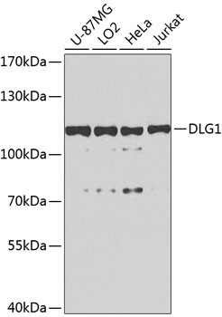
Figure 1. Western blot analysis of SAP97 using anti-SAP97 antibody (PB9552). Electrophoresis was performed on a 5-20% SDS-PAGE gel at 70V (Stacking gel) / 90V (Resolving gel) for 2-3 hours. Lane 1: Rat Lung Tissue Lysate at 50ug, Lane 2: Mouse Lung Tissue Lysate at 50ug, Lane 3: HELA Whole Cell Lysate at 40ug, Lane 4: MM231 Whole Cell Lysate at 40ug, Lane 5: COLO320 Whole Cell Lysate at 40ug, Lane 6: A549 Whole Cell Lysate at 40ug, Lane 7: NIH3T3 Whole Cell Lysate at 40ug. After electrophoresis, proteins were transferred to a nitrocellulose membrane at 150 mA for 50-90 minutes. Blocked the membrane with 5% non-fat milk/TBS for 1.5 hour at RT. The membrane was incubated with rabbit anti-SAP97 antigen affinity purified polyclonal antibody (Catalog # PB9552) at 0.5 microg/mL overnight at 4°C, then washed with TBS-0.1%Tween 3 times with 5 minutes each and probed with a goat anti-rabbit IgG-HRP secondary antibody at a dilution of 1:5000 for 1.5 hour at RT. The signal is developed using an Enhanced Chemiluminescent detection (ECL) kit (Catalog # EK1002) with Tanon 5200 system. A specific band was detected for SAP97 at approximately 130 kDa. The expected band size for SAP97 is at 130 kDa.
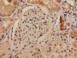
. SAP97 was detected in a paraffin-embedded section of human prostatic cancer tissue. Heat mediated antigen retrieval was performed in EDTA buffer (pH 8.0, epitope retrieval solution). The tissue section was blocked with 10% goat serum. The tissue section was then incubated with 1 microg/ml rabbit anti-SAP97 Antibody (PB9552) overnight at 4°C. Biotinylated goat anti-rabbit IgG was used as secondary antibody and incubated for 30 minutes at 37°C. The tissue section was developed using Strepavidin-Biotin-Complex (SABC) (Catalog # SA1022) with DAB as the chromogen.")
Figure 1. Western blot analysis of SAP97 using anti-SAP97 antibody (PB9552). Electrophoresis was performed on a 5-20% SDS-PAGE gel at 70V (Stacking gel) / 90V (Resolving gel) for 2-3 hours. Lane 1: Rat Lung Tissue Lysate at 50ug, Lane 2: Mouse Lung Tissue Lysate at 50ug, Lane 3: HELA Whole Cell Lysate at 40ug, Lane 4: MM231 Whole Cell Lysate at 40ug, Lane 5: COLO320 Whole Cell Lysate at 40ug, Lane 6: A549 Whole Cell Lysate at 40ug, Lane 7: NIH3T3 Whole Cell Lysate at 40ug. After electrophoresis, proteins were transferred to a nitrocellulose membrane at 150 mA for 50-90 minutes. Blocked the membrane with 5% non-fat milk/TBS for 1.5 hour at RT. The membrane was incubated with rabbit anti-SAP97 antigen affinity purified polyclonal antibody (Catalog # PB9552) at 0.5 microg/mL overnight at 4°C, then washed with TBS-0.1%Tween 3 times with 5 minutes each and probed with a goat anti-rabbit IgG-HRP secondary antibody at a dilution of 1:5000 for 1.5 hour at RT. The signal is developed using an Enhanced Chemiluminescent detection (ECL) kit (Catalog # EK1002) with Tanon 5200 system. A specific band was detected for SAP97 at approximately 130 kDa. The expected band size for SAP97 is at 130 kDa.
Anti-SAP97/DLG1 Antibody Picoband(r)
PB9552-CARRIER-FREE
ApplicationsWestern Blot, ImmunoHistoChemistry
Product group Antibodies
ReactivityHuman, Mouse, Rat
TargetDLG1
Overview
- SupplierBoster Bio
- Product NameAnti-SAP97/DLG1 Antibody Picoband(r)
- Delivery Days Customer9
- Application Supplier NoteTested Species: In-house tested species with positive results. By Heat: Boiling the paraffin sections in 10mM citrate buffer, pH6.0, for 20mins is required for the staining of formalin/paraffin sections. Other applications have not been tested. Optimal dilutions should be determined by end users.
- ApplicationsWestern Blot, ImmunoHistoChemistry
- CertificationResearch Use Only
- ClonalityPolyclonal
- Concentration500 ug/ml
- Gene ID1739
- Target nameDLG1
- Target descriptiondiscs large MAGUK scaffold protein 1
- Target synonymsDLGH1, SAP-97, SAP97, hdlg, disks large homolog 1, dJ1061C18.1.1, discs large homolog 1, scribble cell polarity complex component, presynaptic protein SAP97, synapse-associated protein 97
- HostRabbit
- IsotypeIgG
- Protein IDQ12959
- Protein NameDisks large homolog 1
- Scientific DescriptionBoster Bio Anti-SAP97/DLG1 Antibody Picoband® catalog # PB9552. Tested in IHC, WB applications. This antibody reacts with Human, Mouse, Rat. The brand Picoband indicates this is a premium antibody that guarantees superior quality, high affinity, and strong signals with minimal background in Western blot applications. Only our best-performing antibodies are designated as Picoband, ensuring unmatched performance.
- ReactivityHuman, Mouse, Rat
- Storage Instruction-20°C,2°C to 8°C
- UNSPSC12352203
Related products
Product group Antibodies
DLG1 AntibodyCSB-PA614264LA01HU
ApplicationsImmunoFluorescence, ELISA, ImmunoHistoChemistry
ReactivityHuman
TargetDLG1
- SizePrice
Product group Antibodies
Anti-SAP97 AntibodyA16227
ApplicationsImmunoFluorescence, Western Blot, ImmunoCytoChemistry
ReactivityHuman, Mouse, Rat
- SizePrice
Product group Antibodies

DLG1 / SAP97 AntibodyLS-C747534
ApplicationsWestern Blot
ReactivityHuman, Mouse
TargetDLG1
- SizePrice
Product group Antibodies

Goat anti-DLG1EB08001
ApplicationsFlow Cytometry, ImmunoFluorescence, ELISA
ReactivityHuman, Mouse, Rat
TargetDLG1
- SizePrice
![Various whole cell extracts (30 μg) was separated by 7.5% SDS-PAGE, and the membrane was blotted with SAP97 antibody [GT31] (GTX641123) diluted at 1:1000. The HRP-conjugated anti-mouse IgG antibody (GTX213111-01) was used to detect the primary antibody, and the signal was developed with Trident ECL plus-Enhanced.](https://www.genetex.com/upload/website/prouct_img/normal/GTX641123/GTX641123_45558_20241011_WB_24101600_452.webp)
Product group Antibodies
SAP97 antibody [GT31]GTX641123
ApplicationsWestern Blot
ReactivityHuman, Mouse, Rat
TargetDLG1
- SizePrice
Product group Antibodies
MPP3/SAP102 Polyclonal AntibodyBS-11315R
ApplicationsImmunoFluorescence, Western Blot, ELISA, ImmunoCytoChemistry, ImmunoHistoChemistry, ImmunoHistoChemistry Frozen, ImmunoHistoChemistry Paraffin
ReactivityBovine, Canine, Chicken, Equine, Human, Mouse, Porcine, Rabbit, Rat
TargetDLG1
- SizePrice
Product group Antibodies
Anti-DLG1 Antibody144-08542
ApplicationsWestern Blot, ImmunoHistoChemistry
ReactivityHuman, Mouse, Rat
TargetDLG1
- SizePrice