


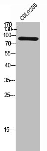
Figure 1. Western blot analysis of SEC23B using anti-SEC23B antibody (A05847-3). Electrophoresis was performed on a 5-20% SDS-PAGE gel at 70V (Stacking gel) / 90V (Resolving gel) for 2-3 hours. The sample well of each lane was loaded with 30ug of sample under reducing conditions. Lane 1: human Raji whole cell lysates, Lane 2: human HepG2 whole cell lysates, Lane 3: human PC-3 whole cell lysates, Lane 4: human A549 whole cell lysates, Lane 5: human THP-1 whole cell lysates, Lane 6: human HL-60 whole cell lysates, Lane 7: rat liver tissue lysates, Lane 8: rat stomach tissue lysates, Lane 9: mouse liver tissue lysates, Lane 10: mouse RAW264.7 whole cell lysates. After Electrophoresis, proteins were transferred to a Nitrocellulose membrane at 150mA for 50-90 minutes. Blocked the membrane with 5% Non-fat Milk/ TBS for 1.5 hour at RT. The membrane was incubated with rabbit anti-SEC23B antigen affinity purified polyclonal antibody (Catalog # A05847-3) at 0.25 microg/mL overnight at 4°C, then washed with TBS-0.1%Tween 3 times with 5 minutes each and probed with a goat anti-rabbit IgG-HRP secondary antibody at a dilution of 1:5000 for 1.5 hour at RT. The signal is developed using an Enhanced Chemiluminescent detection (ECL) kit (Catalog # EK1002) with Tanon 5200 system. A specific band was detected for SEC23B at approximately 86KD. The expected band size for SEC23B is at 86KD.
. Overlay histogram showing SiHa cells stained with A05847-3 (Blue line). To facilitate intracellular staining, cells were fixed with 4% paraformaldehyde and permeabilized with permeabilization buffer. The cells were blocked with 10% normal goat serum. And then incubated with rabbit anti-SEC23B Antibody (A05847-3, 1microg/1x106 cells) for 30 min at 20°C. DyLight®488 conjugated goat anti-rabbit IgG (BA1127, 5-10microg/1x106 cells) was used as secondary antibody for 30 minutes at 20°C. Isotype control antibody (Green line) was rabbit IgG (1microg/1x106) used under the same conditions. Unlabelled sample without incubation with primary antibody and secondary antibody (Red line) was used as a blank control.")
Figure 1. Western blot analysis of SEC23B using anti-SEC23B antibody (A05847-3). Electrophoresis was performed on a 5-20% SDS-PAGE gel at 70V (Stacking gel) / 90V (Resolving gel) for 2-3 hours. The sample well of each lane was loaded with 30ug of sample under reducing conditions. Lane 1: human Raji whole cell lysates, Lane 2: human HepG2 whole cell lysates, Lane 3: human PC-3 whole cell lysates, Lane 4: human A549 whole cell lysates, Lane 5: human THP-1 whole cell lysates, Lane 6: human HL-60 whole cell lysates, Lane 7: rat liver tissue lysates, Lane 8: rat stomach tissue lysates, Lane 9: mouse liver tissue lysates, Lane 10: mouse RAW264.7 whole cell lysates. After Electrophoresis, proteins were transferred to a Nitrocellulose membrane at 150mA for 50-90 minutes. Blocked the membrane with 5% Non-fat Milk/ TBS for 1.5 hour at RT. The membrane was incubated with rabbit anti-SEC23B antigen affinity purified polyclonal antibody (Catalog # A05847-3) at 0.25 microg/mL overnight at 4°C, then washed with TBS-0.1%Tween 3 times with 5 minutes each and probed with a goat anti-rabbit IgG-HRP secondary antibody at a dilution of 1:5000 for 1.5 hour at RT. The signal is developed using an Enhanced Chemiluminescent detection (ECL) kit (Catalog # EK1002) with Tanon 5200 system. A specific band was detected for SEC23B at approximately 86KD. The expected band size for SEC23B is at 86KD.
Anti-SEC23B Antibody Picoband(r)
A05847-3-CARRIER-FREE
ApplicationsFlow Cytometry, Western Blot, ELISA
Product group Antibodies
ReactivityHuman, Mouse, Rat
TargetSEC23B
Overview
- SupplierBoster Bio
- Product NameAnti-SEC23B Antibody Picoband(r)
- Delivery Days Customer9
- ApplicationsFlow Cytometry, Western Blot, ELISA
- CertificationResearch Use Only
- ClonalityPolyclonal
- Concentration500 ug/ml
- Gene ID10483
- Target nameSEC23B
- Target descriptionSEC23 homolog B, COPII component
- Target synonymsCDA-II, CDAII, CDAN2, CWS7, HEMPAS, hSec23B, protein transport protein Sec23B, SEC23 homolog B, COPII coat complex component, SEC23 homolog B, coat complex II component, SEC23-like protein B, SEC23-related protein B, transport protein SEC23B
- HostRabbit
- IsotypeIgG
- Protein IDQ15437
- Protein NameProtein transport protein Sec23B
- Scientific DescriptionBoster Bio Anti-SEC23B Antibody Picoband® catalog # A05847-3. Tested in ELISA, Flow Cytometry, WB applications. This antibody reacts with Human, Mouse, Rat. The brand Picoband indicates this is a premium antibody that guarantees superior quality, high affinity, and strong signals with minimal background in Western blot applications. Only our best-performing antibodies are designated as Picoband, ensuring unmatched performance.
- ReactivityHuman, Mouse, Rat
- Storage Instruction-20°C,2°C to 8°C
- UNSPSC12352203
Related products
Product group Antibodies
Anti-SEC23B Antibody144-60929
ApplicationsWestern Blot
ReactivityHuman
TargetSEC23B
- SizePrice
Product group Antibodies
SEC23B AntibodyCSB-PA006552
ApplicationsWestern Blot, ELISA
ReactivityHuman, Mouse
TargetSEC23B
- SizePrice
Product group Antibodies
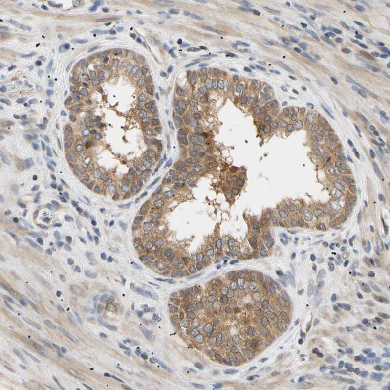
Anti-SEC23B AntibodyHPA008216
ApplicationsImmunoCytoChemistry, ImmunoHistoChemistry
ReactivityHuman
TargetSEC23B
- SizePrice
Product group Antibodies
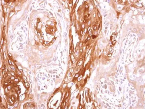
SEC23B antibody [N1N2], N-termGTX119862
ApplicationsWestern Blot, ImmunoHistoChemistry, ImmunoHistoChemistry Paraffin
ReactivityHuman
TargetSEC23B
- SizePrice
Product group Antibodies

SEC23B AntibodyLS-C750102
ApplicationsWestern Blot
ReactivityHuman
TargetSEC23B
- SizePrice
Product group Antibodies
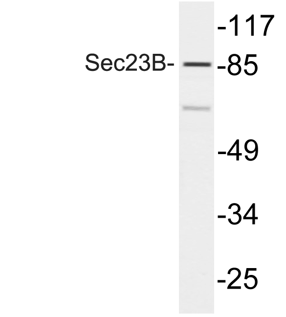
Anti-Sec23B AntibodyA98755
ApplicationsWestern Blot, ELISA
ReactivityHuman, Mouse
- SizePrice