


Immunofluorescent staining of human cell line U-2 OS shows localization to vesicles.

Immunofluorescent staining of human cell line U-2 OS shows localization to vesicles.
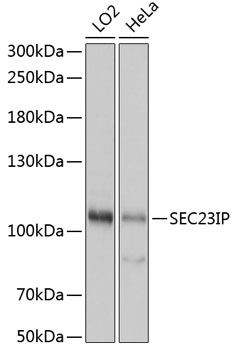
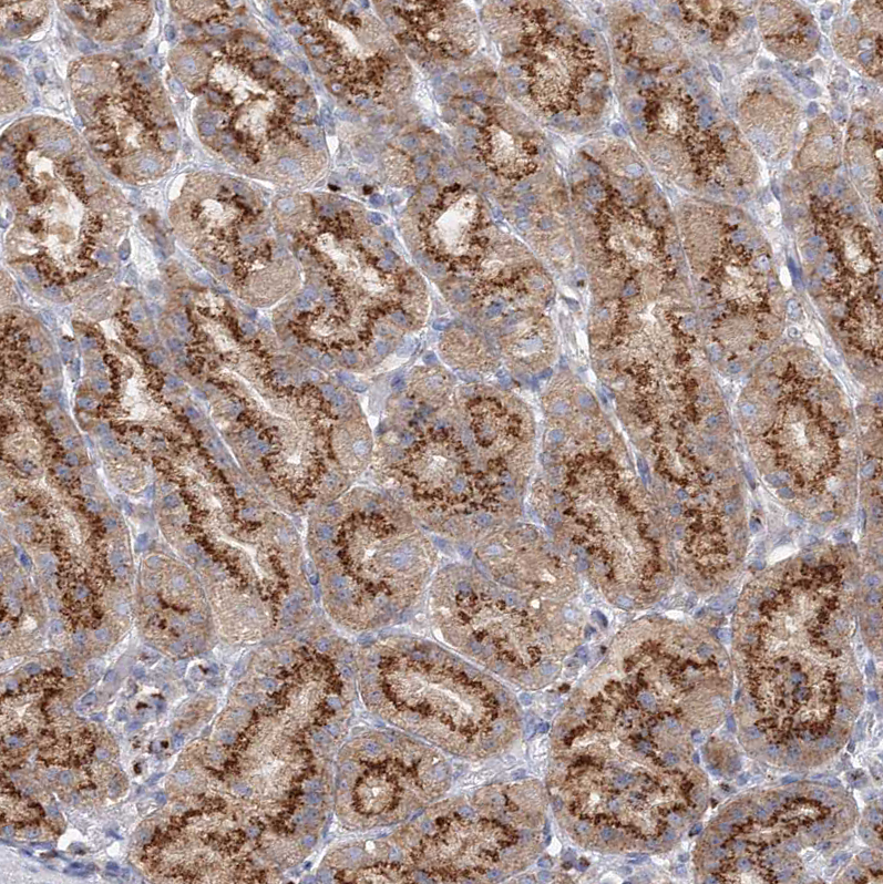
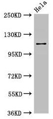
Anti-SEC23IP Antibody
HPA043305
ApplicationsWestern Blot, ImmunoCytoChemistry
Product group Antibodies
ReactivityHuman
TargetSEC23IP
Overview
- SupplierAtlas Antibodies
- Product NameAnti-SEC23IP Antibody
- Delivery Days Customer4
- ApplicationsWestern Blot, ImmunoCytoChemistry
- CertificationResearch Use Only
- ClonalityPolyclonal
- ConjugateUnconjugated
- Gene ID11196
- Target nameSEC23IP
- Target descriptionSEC23 interacting protein
- Target synonymsMSTP053, P125, P125A, iPLA1A, iPLA1beta, SEC23-interacting protein, intracellular phospholipase A1 beta
- HostRabbit
- IsotypeIgG
- Protein IDQ9Y6Y8
- Protein NameSEC23-interacting protein
- Scientific DescriptionRecombinant Protein Epitope Signature Tag (PrEST) antigen sequence
- ReactivityHuman
- Storage Instruction-20°C,2°C to 8°C
- UNSPSC41116161
Datasheet
MSDS
Related products
Product group Antibodies
Anti-SEC23IP AntibodyA92461
ApplicationsImmunoFluorescence, Western Blot, ImmunoCytoChemistry, ImmunoHistoChemistry
- SizePrice
Product group Antibodies
SEC23IP Polyclonal AntibodyCAC12968
ApplicationsImmunoFluorescence, Western Blot, ELISA, ImmunoHistoChemistry
TargetSEC23IP
- SizePrice
Product group Antibodies
Anti-SEC23IP Antibody144-61035
ApplicationsWestern Blot
TargetSEC23IP
- SizePrice
Product group Antibodies
SEC23IP antibodyGTX115597
ApplicationsImmunoFluorescence, Western Blot, ImmunoCytoChemistry, ImmunoHistoChemistry, ImmunoHistoChemistry Paraffin
TargetSEC23IP
- SizePrice
Product group Antibodies

SEC23IP / p125 AntibodyLS-C750368
ApplicationsWestern Blot
TargetSEC23IP
- SizePrice
Product group Antibodies
Anti-SEC23IP-25ulHPA038403
ApplicationsWestern Blot, ImmunoCytoChemistry, ImmunoHistoChemistry
ReactivityHuman, Mouse, Rat
- SizePrice
Product group Antibodies
SEC23IP AntibodyCSB-PA896767LA01HU
ApplicationsImmunoFluorescence, Western Blot, ELISA, ImmunoHistoChemistry
ReactivityHuman
TargetSEC23IP
- SizePrice

Product group Antibodies
Anti-SEC23IP Antibody Picoband(r)A09955-1-CARRIER-FREE
ApplicationsFlow Cytometry, ImmunoFluorescence, Western Blot, ELISA, ImmunoCytoChemistry, ImmunoHistoChemistry
TargetSEC23IP
- SizePrice