


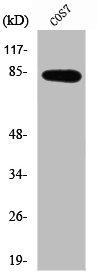
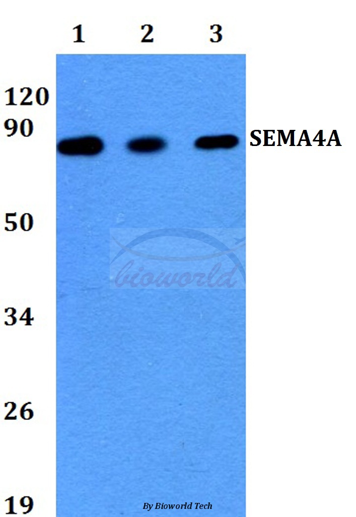
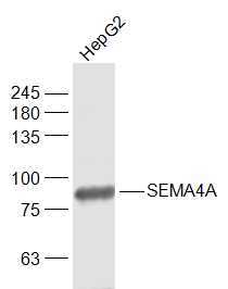
Figure 1. Western blot analysis of SEMA4A using anti-SEMA4A antibody (A06779-3). Electrophoresis was performed on a 5-20% SDS-PAGE gel at 70V (Stacking gel) / 90V (Resolving gel) for 2-3 hours. The sample well of each lane was loaded with 30 ug of sample under reducing conditions. Lane 1: human Hela whole cell lysates, Lane 2: human Jurkat whole cell lysates, Lane 3: human RT4 whole cell lysates, Lane 4: rat thymus tissue lysates, Lane 5: rat PC-12 whole cell lysates, Lane 6: mouse thymus tissue lysates, Lane 7: mouse ANA-1 tissue lysates. After electrophoresis, proteins were transferred to a nitrocellulose membrane at 150 mA for 50-90 minutes. Blocked the membrane with 5% non-fat milk/TBS for 1.5 hour at RT. The membrane was incubated with rabbit anti-SEMA4A antigen affinity purified polyclonal antibody (Catalog # A06779-3) at 0.5 microg/mL overnight at 4°C, then washed with TBS-0.1%Tween 3 times with 5 minutes each and probed with a goat anti-rabbit IgG-HRP secondary antibody at a dilution of 1:5000 for 1.5 hour at RT. The signal is developed using an Enhanced Chemiluminescent detection (ECL) kit (Catalog # EK1002) with Tanon 5200 system. A specific band was detected for SEMA4A at approximately 105 kDa. The expected band size for SEMA4A is at 84,105 kDa.
. Overlay histogram showing JK cells stained with A06779-3 (Blue line). The cells were fixed with 4% paraformaldehyde and blocked with 10% normal goat serum. And then incubated with rabbit anti-SEMA4A Antibody (A06779-3, 1 microg/1x106 cells) for 30 min at 20°C. DyLight®488 conjugated goat anti-rabbit IgG (BA1127, 5-10 microg/1x106 cells) was used as secondary antibody for 30 minutes at 20°C. Isotype control antibody (Green line) was rabbit IgG (1 microg/1x106) used under the same conditions. Unlabelled sample (Red line) was also used as a control.")
Figure 1. Western blot analysis of SEMA4A using anti-SEMA4A antibody (A06779-3). Electrophoresis was performed on a 5-20% SDS-PAGE gel at 70V (Stacking gel) / 90V (Resolving gel) for 2-3 hours. The sample well of each lane was loaded with 30 ug of sample under reducing conditions. Lane 1: human Hela whole cell lysates, Lane 2: human Jurkat whole cell lysates, Lane 3: human RT4 whole cell lysates, Lane 4: rat thymus tissue lysates, Lane 5: rat PC-12 whole cell lysates, Lane 6: mouse thymus tissue lysates, Lane 7: mouse ANA-1 tissue lysates. After electrophoresis, proteins were transferred to a nitrocellulose membrane at 150 mA for 50-90 minutes. Blocked the membrane with 5% non-fat milk/TBS for 1.5 hour at RT. The membrane was incubated with rabbit anti-SEMA4A antigen affinity purified polyclonal antibody (Catalog # A06779-3) at 0.5 microg/mL overnight at 4°C, then washed with TBS-0.1%Tween 3 times with 5 minutes each and probed with a goat anti-rabbit IgG-HRP secondary antibody at a dilution of 1:5000 for 1.5 hour at RT. The signal is developed using an Enhanced Chemiluminescent detection (ECL) kit (Catalog # EK1002) with Tanon 5200 system. A specific band was detected for SEMA4A at approximately 105 kDa. The expected band size for SEMA4A is at 84,105 kDa.
Anti-SEMA4A Antibody Picoband(r)
A06779-3-CARRIER-FREE
ApplicationsFlow Cytometry, Western Blot, ELISA
Product group Antibodies
ReactivityHuman, Mouse, Rat
TargetSEMA4A
Overview
- SupplierBoster Bio
- Product NameAnti-SEMA4A Antibody Picoband(r)
- Delivery Days Customer9
- ApplicationsFlow Cytometry, Western Blot, ELISA
- CertificationResearch Use Only
- ClonalityPolyclonal
- Concentration500 ug/ml
- Gene ID64218
- Target nameSEMA4A
- Target descriptionsemaphorin 4A
- Target synonymsCORD10, RP35, SEMAB, SEMB, semaphorin-4A, sema B, sema domain, immunoglobulin domain (Ig), transmembrane domain (TM) and short cytoplasmic domain, (semaphorin) 4A, semaphorin-B
- HostRabbit
- IsotypeIgG
- Protein IDQ9H3S1
- Protein NameSemaphorin-4A
- Scientific DescriptionBoster Bio Anti-SEMA4A Antibody Picoband® catalog # A06779-3. Tested in ELISA, WB, Flow Cytometry applications. This antibody reacts with Human, Mouse, Rat. The brand Picoband indicates this is a premium antibody that guarantees superior quality, high affinity, and strong signals with minimal background in Western blot applications. Only our best-performing antibodies are designated as Picoband, ensuring unmatched performance.
- ReactivityHuman, Mouse, Rat
- Storage Instruction-20°C,2°C to 8°C
- UNSPSC12352203
Related products
Product group Antibodies
SEMA4A AntibodyCSB-PA004061
ApplicationsImmunoFluorescence, Western Blot, ELISA, ImmunoHistoChemistry
ReactivityHuman, Monkey, Mouse, Rat
TargetSEMA4A
- SizePrice
Product group Antibodies
ApplicationsImmunoFluorescence, Western Blot, ImmunoHistoChemistry
ReactivityHuman, Mouse, Rat
- SizePrice

Product group Antibodies
Anti-SEMA4A AntibodyHPA069136
ApplicationsImmunoCytoChemistry, ImmunoHistoChemistry
ReactivityHuman
TargetSEMA4A
- SizePrice
Product group Antibodies

SEMA4A / Semaphorin 4A AntibodyLS-C405328
ApplicationsELISA, ImmunoHistoChemistry
ReactivityHuman
TargetSEMA4A
- SizePrice
Product group Antibodies
SEMA4A Polyclonal AntibodyBS-7519R
ApplicationsImmunoFluorescence, Western Blot, ELISA, ImmunoCytoChemistry, ImmunoHistoChemistry, ImmunoHistoChemistry Frozen, ImmunoHistoChemistry Paraffin
ReactivityBovine, Canine, Human, Mouse, Porcine, Rabbit, Rat
TargetSEMA4A
- SizePrice
Product group Antibodies
SEMA4A antibody [C1C3]GTX109538
ApplicationsWestern Blot, ImmunoHistoChemistry, ImmunoHistoChemistry Paraffin
ReactivityHuman
TargetSEMA4A
- SizePrice
Product group Antibodies
Anti-SEMA4A (N-term) Antibody102-22446
ApplicationsWestern Blot
TargetSEMA4A
- SizePrice