

Anti-SEPHS1 Antibody
A31394
ApplicationsWestern Blot, ImmunoHistoChemistry
Product group Antibodies
ReactivityHuman, Mouse, Rat
Overview
- SupplierAntibodies.com
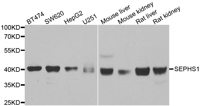
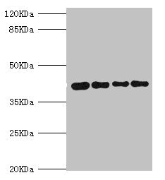
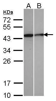
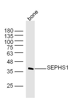
- Product NameAnti-SEPHS1 Antibody
- Delivery Days Customer7
- ApplicationsWestern Blot, ImmunoHistoChemistry
- CertificationResearch Use Only
- ClonalityPolyclonal
- ConjugateUnconjugated
- Estimated Purity>95%
- HostRabbit
- Scientific DescriptionRabbit polyclonal antibody to SEPHS1
- ReactivityHuman, Mouse, Rat
- UNSPSC12352203
Related products
Product group Antibodies
SEPHS1 AntibodyCSB-PA021015ESR2HU
ApplicationsWestern Blot, ELISA, ImmunoHistoChemistry
ReactivityHuman, Mouse
TargetSEPHS1
- SizePrice

Product group Antibodies
Anti-Selenophosphate synthetase 1/SEPHS1 Antibody Picoband(r)A10102-1-CARRIER-FREE
ApplicationsWestern Blot, ImmunoHistoChemistry
ReactivityHuman, Mouse, Rat
TargetSEPHS1
- SizePrice
Product group Antibodies

SEPHS1 / SPS AntibodyLS-C831019
ApplicationsELISA, ImmunoHistoChemistry
ReactivityHuman, Mouse
TargetSEPHS1
- SizePrice

Product group Antibodies
Anti-SEPHS1 AntibodyHPA037645
ApplicationsWestern Blot, ImmunoHistoChemistry
ReactivityHuman, Mouse, Rat
TargetSEPHS1
- SizePrice
Product group Antibodies
ApplicationsImmunoFluorescence, Western Blot, ImmunoCytoChemistry, ImmunoHistoChemistry, ImmunoHistoChemistry Paraffin
ReactivityHuman
TargetSEPHS1
- SizePrice
Product group Antibodies
Anti-SEPHS1 Antibody144-60322
ApplicationsImmunoFluorescence, Western Blot
ReactivityHuman, Mouse, Rat
TargetSEPHS1
- SizePrice
Product group Antibodies
SEPHS1 Polyclonal AntibodyBS-19627R
ApplicationsImmunoFluorescence, Western Blot, ELISA, ImmunoCytoChemistry, ImmunoHistoChemistry, ImmunoHistoChemistry Frozen, ImmunoHistoChemistry Paraffin
ReactivityBovine, Canine, Chicken, Equine, Human, Mouse, Porcine, Rabbit, Rat
TargetSEPHS1
- SizePrice