


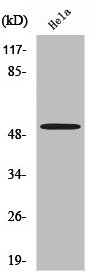
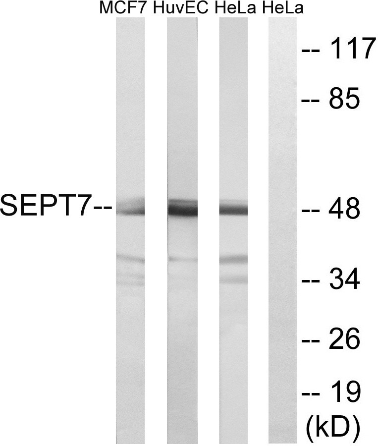
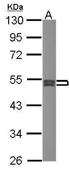
Figure 1. Western blot analysis of SEPT7/SEPTIN7 using anti-SEPT7/SEPTIN7 antibody (A30759-1). Electrophoresis was performed on a 5-20% SDS-PAGE gel at 70V (Stacking gel) / 90V (Resolving gel) for 2-3 hours. The sample well of each lane was loaded with 30 ug of sample under reducing conditions. Lane 1: human Hela whole cell lysates, Lane 2: human MCF-7 whole cell lysates, Lane 3: human HepG2 whole cell lysates, Lane 4: human A431 whole cell lysates, Lane 5: human U20S whole cell lysates, Lane 6: human A375 whole cell lysates, Lane 7: rat testis tissue lysates, Lane 8: rat brain tissue lysates, Lane 9: rat stomach tissue lysates, Lane 10: rat PC-12 whole cell lysates, Lane 11: mouse brain tissue lysates, Lane 12: mouse Neuro-2a whole cell lysates. red to a nitrocellulose membrane at 150 mA for 50-90 minutes. Blocked the membrane with 5% non-fat milk/TBS for 1.5 hour at RT. The membrane was incubated with rabbit anti-SEPT7/SEPTIN7 antigen affinity purified polyclonal antibody (Catalog # A30759-1) at 0.5 microg/mL overnight at 4°C, then washed with TBS-0.1%Tween 3 times with 5 minutes each and probed with a goat anti-rabbit IgG-HRP secondary antibody at a dilution of 1:5000 for 1.5 hour at RT. The signal is developed using an Enhanced Chemiluminescent detection (ECL) kit (Catalog # EK1002) with Tanon 5200 system. A specific band was detected for SEPT7/SEPTIN7 at approximately 51 kDa. The expected band size for SEPT7/SEPTIN7 is at 51 kDa.
. SEPT7/SEPTIN7 was detected in a paraffin-embedded section of human colorectal adenocarcinoma tissue. Heat mediated antigen retrieval was performed in EDTA buffer (pH 8.0, epitope retrieval solution). The tissue section was blocked with 10% goat serum. The tissue section was then incubated with 2 microg/ml rabbit anti-SEPT7/SEPTIN7 Antibody (A30759-1) overnight at 4°C. Peroxidase Conjugated Goat Anti-rabbit IgG was used as secondary antibody and incubated for 30 minutes at 37°C. The tissue section was developed using HRP Conjugated Rabbit IgG Super Vision Assay Kit (Catalog # SV0002) with DAB as the chromogen.")
. SEPT7/SEPTIN7 was detected in a paraffin-embedded section of human ovarian cancer tissue. Heat mediated antigen retrieval was performed in EDTA buffer (pH 8.0, epitope retrieval solution). The tissue section was blocked with 10% goat serum. The tissue section was then incubated with 2 microg/ml rabbit anti-SEPT7/SEPTIN7 Antibody (A30759-1) overnight at 4°C. Peroxidase Conjugated Goat Anti-rabbit IgG was used as secondary antibody and incubated for 30 minutes at 37°C. The tissue section was developed using HRP Conjugated Rabbit IgG Super Vision Assay Kit (Catalog # SV0002) with DAB as the chromogen.")
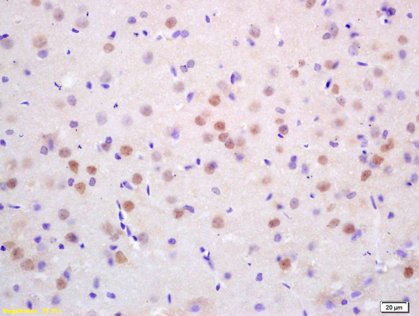
. SEPT7/SEPTIN7 was detected in a paraffin-embedded section of rat brain tissue. Heat mediated antigen retrieval was performed in EDTA buffer (pH 8.0, epitope retrieval solution). The tissue section was blocked with 10% goat serum. The tissue section was then incubated with 2 microg/ml rabbit anti-SEPT7/SEPTIN7 Antibody (A30759-1) overnight at 4°C. Peroxidase Conjugated Goat Anti-rabbit IgG was used as secondary antibody and incubated for 30 minutes at 37°C. The tissue section was developed using HRP Conjugated Rabbit IgG Super Vision Assay Kit (Catalog # SV0002) with DAB as the chromogen.")
. SEPT7/SEPTIN7 was detected in a paraffin-embedded section of human ovarian cancer tissue. Heat mediated antigen retrieval was performed in EDTA buffer (pH 8.0, epitope retrieval solution). The tissue section was blocked with 10% goat serum. The tissue section was then incubated with 5 microg/mL rabbit anti-SEPT7/SEPTIN7 Antibody (A30759-1) overnight at 4°C. Cy3 Conjugated Goat Anti-Rabbit IgG (BA1032) was used as secondary antibody at 1:500 dilution and incubated for 30 minutes at 37°C. The section was counterstained with DAPI. Visualize using a fluorescence microscope and filter sets appropriate for the label used.")
. Overlay histogram showing Hela cells stained with A30759-1 (Blue line). To facilitate intracellular staining, cells were fixed with 4% paraformaldehyde and permeabilized with permeabilization buffer. The cells were blocked with 10% normal goat serum. And then incubated with rabbit anti-SEPT7/SEPTIN7 Antibody (A30759-1, 1 microg/1x106 cells) for 30 min at 20°C. DyLight®488 conjugated goat anti-rabbit IgG (BA1127, 5-10 microg/1x106 cells) was used as secondary antibody for 30 minutes at 20°C. Isotype control antibody (Green line) was rabbit IgG (1 microg/1x106) used under the same conditions. Unlabelled sample without incubation with primary antibody and secondary antibody (Red line) was used as a blank control.")
Figure 1. Western blot analysis of SEPT7/SEPTIN7 using anti-SEPT7/SEPTIN7 antibody (A30759-1). Electrophoresis was performed on a 5-20% SDS-PAGE gel at 70V (Stacking gel) / 90V (Resolving gel) for 2-3 hours. The sample well of each lane was loaded with 30 ug of sample under reducing conditions. Lane 1: human Hela whole cell lysates, Lane 2: human MCF-7 whole cell lysates, Lane 3: human HepG2 whole cell lysates, Lane 4: human A431 whole cell lysates, Lane 5: human U20S whole cell lysates, Lane 6: human A375 whole cell lysates, Lane 7: rat testis tissue lysates, Lane 8: rat brain tissue lysates, Lane 9: rat stomach tissue lysates, Lane 10: rat PC-12 whole cell lysates, Lane 11: mouse brain tissue lysates, Lane 12: mouse Neuro-2a whole cell lysates. red to a nitrocellulose membrane at 150 mA for 50-90 minutes. Blocked the membrane with 5% non-fat milk/TBS for 1.5 hour at RT. The membrane was incubated with rabbit anti-SEPT7/SEPTIN7 antigen affinity purified polyclonal antibody (Catalog # A30759-1) at 0.5 microg/mL overnight at 4°C, then washed with TBS-0.1%Tween 3 times with 5 minutes each and probed with a goat anti-rabbit IgG-HRP secondary antibody at a dilution of 1:5000 for 1.5 hour at RT. The signal is developed using an Enhanced Chemiluminescent detection (ECL) kit (Catalog # EK1002) with Tanon 5200 system. A specific band was detected for SEPT7/SEPTIN7 at approximately 51 kDa. The expected band size for SEPT7/SEPTIN7 is at 51 kDa.
Anti-SEPT7/SEPTIN7 Antibody Picoband(r)
A30759-1-CARRIER-FREE
ApplicationsFlow Cytometry, ImmunoFluorescence, Western Blot, ELISA, ImmunoHistoChemistry
Product group Antibodies
TargetSEPTIN7
Overview
- SupplierBoster Bio
- Product NameAnti-SEPT7/SEPTIN7 Antibody Picoband(r)
- Delivery Days Customer9
- ApplicationsFlow Cytometry, ImmunoFluorescence, Western Blot, ELISA, ImmunoHistoChemistry
- CertificationResearch Use Only
- ClonalityPolyclonal
- Concentration500 ug/ml
- Gene ID989
- Target nameSEPTIN7
- Target descriptionseptin 7
- Target synonymsCDC10, CDC3, NBLA02942, SEPT7, SEPT7A, Septin-7, septin-7, CDC10 (cell division cycle 10, S. cerevisiae, homolog), CDC10 protein homolog
- HostRabbit
- IsotypeIgG
- Protein IDQ16181
- Protein NameSeptin-7
- Scientific DescriptionBoster Bio Anti-SEPT7/SEPTIN7 Antibody Picoband® catalog # A30759-1. Tested in ELISA, Flow Cytometry, IF, IHC, WB applications. This antibody reacts with Human, Mouse, Rat. The brand Picoband indicates this is a premium antibody that guarantees superior quality, high affinity, and strong signals with minimal background in Western blot applications. Only our best-performing antibodies are designated as Picoband, ensuring unmatched performance.
- Storage Instruction-20°C,2°C to 8°C
- UNSPSC12352203
Related products
Product group Antibodies
SEPT7 AntibodyCSB-PA004069
ApplicationsWestern Blot, ELISA, ImmunoHistoChemistry
ReactivityHuman, Mouse, Rat
TargetSEPTIN7
- SizePrice
Product group Antibodies
Anti-SEPT7 AntibodyA97240
ApplicationsWestern Blot, ELISA
ReactivityHuman, Mouse, Rat
- SizePrice
Product group Antibodies

SEPT7 / Septin 7 AntibodyLS-C770522
ApplicationsWestern Blot, ELISA, ImmunoHistoChemistry
ReactivityHuman, Mouse, Rat
TargetSEPTIN7
- SizePrice
Product group Antibodies

Goat anti-SEPT7EB08933
ApplicationsWestern Blot, ELISA
ReactivityBovine, Canine, Equine, Human, Mouse, Porcine, Rat
TargetSEPTIN7
- SizePrice

Product group Antibodies
Anti-SEPT7 AntibodyHPA029524
ApplicationsWestern Blot, ImmunoCytoChemistry, ImmunoHistoChemistry
ReactivityHuman, Mouse, Rat
TargetSEPTIN7
- SizePrice
Product group Antibodies
45176 Polyclonal AntibodyCAC10573
ApplicationsWestern Blot, ELISA, ImmunoHistoChemistry
ReactivityMouse
TargetSEPTIN7
- SizePrice
Product group Antibodies
SEPT7 antibody [N3C3]GTX101533
ApplicationsWestern Blot, ImmunoHistoChemistry, ImmunoHistoChemistry Paraffin
ReactivityHuman, Zebra Fish
TargetSEPTIN7
- SizePrice
Product group Antibodies
Anti-Septin 7 Antibody102-26123
ApplicationsWestern Blot
TargetSEPTIN7
- SizePrice
Product group Antibodies
CDC10 Polyclonal AntibodyBS-7741R
ApplicationsImmunoFluorescence, Western Blot, ELISA, ImmunoCytoChemistry, ImmunoHistoChemistry, ImmunoHistoChemistry Frozen, ImmunoHistoChemistry Paraffin
ReactivityBovine, Human, Mouse, Rat, Zebra Fish
TargetSEPTIN7
- SizePrice