


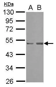
Western blot analysis of SETD7 in (1) Jurkat cell lysate; (2) HeLa cell lysate.
Western blot analysis of SETD7 in (1) Jurkat cell lysate; (2) HeLa cell lysate.
Anti-SETD7/Set7 Rabbit Monoclonal Antibody
M02793
ApplicationsImmunoPrecipitation, Western Blot
Product group Antibodies
ReactivityHuman, Mouse, Rat
TargetSETD7
Overview
- SupplierBoster Bio
- Product NameAnti-SETD7/Set7 Rabbit Monoclonal Antibody
- Delivery Days Customer9
- ApplicationsImmunoPrecipitation, Western Blot
- CertificationResearch Use Only
- ClonalityMonoclonal
- Clone IDABA-19
- Gene ID80854
- Target nameSETD7
- Target descriptionSET domain containing 7, histone lysine methyltransferase
- Target synonymsKMT7, SET7, SET7/9, SET9, histone-lysine N-methyltransferase SETD7, H3-K4-HMTase SETD7, SET domain containing 7, lysine methyltransferase, SET domain containing lysine methyltransferase 7, SET domain-containing protein 7, histone H3-K4 methyltransferase SETD7, histone H3-lysine 4-specific methyltransferase, lysine N-methyltransferase 7
- HostRabbit
- IsotypeIgG
- Protein IDQ8WTS6
- Protein NameHistone-lysine N-methyltransferase SETD7
- Scientific DescriptionBoster Bio Anti-SETD7/Set7 Rabbit Monoclonal Antibody catalog # M02793. Tested in WB, IP applications. This antibody reacts with Human, Mouse, Rat.
- ReactivityHuman, Mouse, Rat
- Storage Instruction-20°C
- UNSPSC12352203
Datasheet
MSDS
Related products

Product group Antibodies
Anti-SETD7 AntibodyA46010
ApplicationsImmunoHistoChemistry
ReactivityHuman
- SizePrice
Product group Antibodies
Anti-SETD7 Antibody144-09985
ApplicationsWestern Blot
ReactivityHuman, Mouse, Rat
TargetSETD7
- SizePrice

Product group Antibodies
ApplicationsWestern Blot
ReactivityHuman
TargetSETD7
- SizePrice
Product group Antibodies

Anti-SETD7 [RAB-C220]AB01713-1.1-BT
ApplicationsFlow Cytometry, ImmunoFluorescence, ImmunoPrecipitation, ChIP Chromatin ImmunoPrecipitation, ELISA
ReactivityHuman
TargetSETD7
- SizePrice
Product group Antibodies
H3-K4-HMTase SETD7 Recombinant AntibodyBSM-61109R
ApplicationsFlow Cytometry, ImmunoPrecipitation, Western Blot
TargetSETD7
- SizePrice
Product group Antibodies
SETD7 AntibodyCSB-PA093769
ApplicationsELISA, ImmunoHistoChemistry
ReactivityHuman, Mouse
TargetSETD7
- SizePrice
Product group Antibodies
SETD7 Polyclonal AntibodyCAC14316
ApplicationsWestern Blot, ELISA, ImmunoHistoChemistry
TargetSETD7
- SizePrice

Product group Antibodies
Anti-SETD7 AntibodyHPA058111
ApplicationsWestern Blot, ImmunoCytoChemistry, ImmunoHistoChemistry
ReactivityHuman
TargetSETD7
- SizePrice
Product group Antibodies
SETD7 antibody [N1C1]GTX117333
ApplicationsWestern Blot
ReactivityHuman
TargetSETD7
- SizePrice