


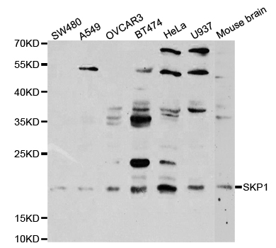
Figure 1. Western blot analysis of SKP1 using anti-SKP1 antibody (A00476-2). Electrophoresis was performed on a 5-20% SDS-PAGE gel at 70V (Stacking gel) / 90V (Resolving gel) for 2-3 hours. The sample well of each lane was loaded with 50ug of sample under reducing conditions. Lane 1: human U-87MG whole cell lysates, Lane 2: human T-47D whole cell lysates, Lane 3: human Caco-2 whole cell lysates, Lane 4: human PC-3 whole cell lysates, Lane 5: human K562 whole cell lysates, Lane 6: rat brain tissue lysates, Lane 7: mouse brain tissue lysates, Lane 8: mouse RAW264.7 whole cell lysates. After Electrophoresis, proteins were transferred to a Nitrocellulose membrane at 150mA for 50-90 minutes. Blocked the membrane with 5% Non-fat Milk/ TBS for 1.5 hour at RT. The membrane was incubated with rabbit anti-SKP1 antigen affinity purified polyclonal antibody (Catalog # A00476-2) at 0.5 microg/mL overnight at 4°C, then washed with TBS-0.1%Tween 3 times with 5 minutes each and probed with a goat anti-rabbit IgG-HRP secondary antibody at a dilution of 1:10000 for 1.5 hour at RT. The signal is developed using an Enhanced Chemiluminescent detection (ECL) kit (Catalog # EK1002) with Tanon 5200 system. A specific band was detected for SKP1 at approximately 19KD. The expected band size for SKP1 is at 19KD.
. SKP1 was detected in paraffin-embedded section of human colon cancer tissues. Heat mediated antigen retrieval was performed in citrate buffer (pH6, epitope retrieval solution) for 20 mins. The tissue section was blocked with 10% goat serum. The tissue section was then incubated with 1microg/ml rabbit anti-SKP1 Antibody (A00476-2) overnight at 4°C. Biotinylated goat anti-rabbit IgG was used as secondary antibody and incubated for 30 minutes at 37°C. The tissue section was developed using Strepavidin-Biotin-Complex (SABC)(Catalog # SA1022) with DAB as the chromogen.")
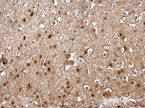
. SKP1 was detected in paraffin-embedded section of mouse brain tissues. Heat mediated antigen retrieval was performed in citrate buffer (pH6, epitope retrieval solution) for 20 mins. The tissue section was blocked with 10% goat serum. The tissue section was then incubated with 1microg/ml rabbit anti-SKP1 Antibody (A00476-2) overnight at 4°C. Biotinylated goat anti-rabbit IgG was used as secondary antibody and incubated for 30 minutes at 37°C. The tissue section was developed using Strepavidin-Biotin-Complex (SABC)(Catalog # SA1022) with DAB as the chromogen.")
. SKP1 was detected in paraffin-embedded section of rat brain tissues. Heat mediated antigen retrieval was performed in citrate buffer (pH6, epitope retrieval solution) for 20 mins. The tissue section was blocked with 10% goat serum. The tissue section was then incubated with 1microg/ml rabbit anti-SKP1 Antibody (A00476-2) overnight at 4°C. Biotinylated goat anti-rabbit IgG was used as secondary antibody and incubated for 30 minutes at 37°C. The tissue section was developed using Strepavidin-Biotin-Complex (SABC)(Catalog # SA1022) with DAB as the chromogen.")
. SKP1 was detected in paraffin-embedded section of rat brain tissues. Heat mediated antigen retrieval was performed in citrate buffer (pH6, epitope retrieval solution) for 20 mins. The tissue section was blocked with 10% goat serum. The tissue section was then incubated with 1microg/ml rabbit anti-SKP1 Antibody (A00476-2) overnight at 4°C. Biotinylated goat anti-rabbit IgG was used as secondary antibody and incubated for 30 minutes at 37°C. The tissue section was developed using Strepavidin-Biotin-Complex (SABC)(Catalog # SA1022) with DAB as the chromogen.")
Figure 1. Western blot analysis of SKP1 using anti-SKP1 antibody (A00476-2). Electrophoresis was performed on a 5-20% SDS-PAGE gel at 70V (Stacking gel) / 90V (Resolving gel) for 2-3 hours. The sample well of each lane was loaded with 50ug of sample under reducing conditions. Lane 1: human U-87MG whole cell lysates, Lane 2: human T-47D whole cell lysates, Lane 3: human Caco-2 whole cell lysates, Lane 4: human PC-3 whole cell lysates, Lane 5: human K562 whole cell lysates, Lane 6: rat brain tissue lysates, Lane 7: mouse brain tissue lysates, Lane 8: mouse RAW264.7 whole cell lysates. After Electrophoresis, proteins were transferred to a Nitrocellulose membrane at 150mA for 50-90 minutes. Blocked the membrane with 5% Non-fat Milk/ TBS for 1.5 hour at RT. The membrane was incubated with rabbit anti-SKP1 antigen affinity purified polyclonal antibody (Catalog # A00476-2) at 0.5 microg/mL overnight at 4°C, then washed with TBS-0.1%Tween 3 times with 5 minutes each and probed with a goat anti-rabbit IgG-HRP secondary antibody at a dilution of 1:10000 for 1.5 hour at RT. The signal is developed using an Enhanced Chemiluminescent detection (ECL) kit (Catalog # EK1002) with Tanon 5200 system. A specific band was detected for SKP1 at approximately 19KD. The expected band size for SKP1 is at 19KD.
Anti-SKP1 Antibody Picoband(r)
A00476-2-CARRIER-FREE
ApplicationsFlow Cytometry, ImmunoFluorescence, Western Blot, ELISA, ImmunoCytoChemistry, ImmunoHistoChemistry
Product group Antibodies
ReactivityHuman, Mouse, Rat
TargetSKP1
Overview
- SupplierBoster Bio
- Product NameAnti-SKP1 Antibody Picoband(r)
- Delivery Days Customer9
- ApplicationsFlow Cytometry, ImmunoFluorescence, Western Blot, ELISA, ImmunoCytoChemistry, ImmunoHistoChemistry
- CertificationResearch Use Only
- ClonalityPolyclonal
- Concentration500 ug/ml
- Gene ID6500
- Target nameSKP1
- Target descriptionS-phase kinase associated protein 1
- Target synonymsEMC19, OCP-II, OCP2, SKP1A, TCEB1L, p19A, S-phase kinase-associated protein 1, OCP-2, RNA polymerase II elongation factor-like protein OCP2, SIII, cyclin A/CDK2-associated p19, cyclin-A/CDK2-associated protein p19, organ of Corti protein 2, organ of Corti protein II, p19skp1, transcription elongation factor B polypeptide 1-like
- HostRabbit
- IsotypeIgG
- Protein IDP63208
- Protein NameS-phase kinase-associated protein 1
- Scientific DescriptionBoster Bio Anti-SKP1 Antibody Picoband® catalog # A00476-2. Tested in ELISA, Flow Cytometry, IF, IHC, ICC, WB applications. This antibody reacts with Human, Mouse, Rat. The brand Picoband indicates this is a premium antibody that guarantees superior quality, high affinity, and strong signals with minimal background in Western blot applications. Only our best-performing antibodies are designated as Picoband, ensuring unmatched performance.
- ReactivityHuman, Mouse, Rat
- Storage Instruction-20°C,2°C to 8°C
- UNSPSC12352203
Related products
Product group Antibodies
Anti-Skp1 p19 AntibodyA30425
ApplicationsWestern Blot, ImmunoHistoChemistry
ReactivityHuman, Mouse, Rat
- SizePrice
Product group Antibodies
Anti-SKP1 Antibody144-02566
ApplicationsWestern Blot, ImmunoHistoChemistry
ReactivityHuman, Mouse
TargetSKP1
- SizePrice
Product group Antibodies
SKP1 Recombinant Antibody, AbBy Fluor-488 ConjugatedBSM-61662R-BF488
ApplicationsFlow Cytometry, Western Blot
ReactivityHuman, Mouse, Rat
TargetSKP1
- SizePrice
Product group Antibodies
SKP1 AntibodyCSB-PA004097
ApplicationsWestern Blot, ELISA, ImmunoHistoChemistry
ReactivityHuman, Monkey, Mouse, Rat
TargetSKP1
- SizePrice
Product group Antibodies
SKP1 Polyclonal AntibodyCAC13932
ApplicationsWestern Blot, ELISA
ReactivityMouse
TargetSKP1
- SizePrice

Product group Antibodies
Anti-SKP1 AntibodyHPA058134
ApplicationsImmunoCytoChemistry
ReactivityHuman
TargetSKP1
- SizePrice
Product group Antibodies
SKP1 antibodyGTX106675
ApplicationsWestern Blot, ImmunoHistoChemistry, ImmunoHistoChemistry Paraffin
ReactivityHuman, Mouse
TargetSKP1
- SizePrice
Product group Antibodies

SKP1 AntibodyLS-C404283
ApplicationsWestern Blot, ELISA, ImmunoHistoChemistry
ReactivityHuman, Mouse, Rat
TargetSKP1
- SizePrice
Product group Antibodies
Anti-SKP1 AntibodyCAB2566
ApplicationsWestern Blot, ELISA
ReactivityHuman
TargetSKP1
- SizePrice