


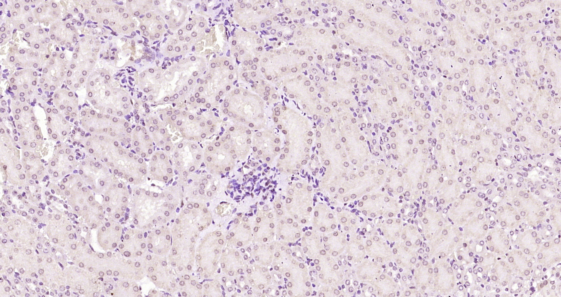
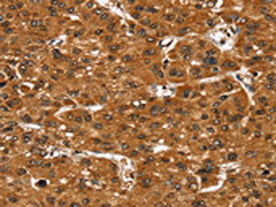
Immunohistochemical staining of human duodenum shows strong nuclear positivity in glandular cells.

Immunohistochemical staining of human duodenum shows strong nuclear positivity in glandular cells.
Anti-SMARCB1 Antibody
HPA019127
ApplicationsWestern Blot, ChIP Chromatin ImmunoPrecipitation, ImmunoHistoChemistry
Product group Antibodies
ReactivityHuman, Mouse, Rat
TargetSMARCB1
Overview
- SupplierAtlas Antibodies
- Product NameAnti-SMARCB1 Antibody
- Delivery Days Customer4
- ApplicationsWestern Blot, ChIP Chromatin ImmunoPrecipitation, ImmunoHistoChemistry
- CertificationResearch Use Only
- ClonalityPolyclonal
- ConjugateUnconjugated
- Gene ID6598
- Target nameSMARCB1
- Target descriptionSWI/SNF related BAF chromatin remodeling complex subunit B1
- Target synonymsBAF47, CSS3, INI-1, INI1, MRD15, PPP1R144, RDT, RTPS1, SNF5, SNF5L1, SWNTS1, Sfh1p, Snr1, hSNFS, SWI/SNF-related matrix-associated actin-dependent regulator of chromatin subfamily B member 1, BRG1-associated factor 47, SNF5 homolog, SWI/SNF related, matrix associated, actin dependent regulator of chromatin, subfamily b, member 1, SWI/SNF-related matrix-associated protein, hSNF5, integrase interactor 1 protein, malignant rhabdoid tumor suppressor, protein phosphatase 1, regulatory subunit 144, sucrose nonfermenting, yeast, homolog-like 1
- HostRabbit
- IsotypeIgG
- Protein IDQ12824
- Protein NameSWI/SNF-related matrix-associated actin-dependent regulator of chromatin subfamily B member 1
- Scientific DescriptionRecombinant Protein Epitope Signature Tag (PrEST) antigen sequence
- ReactivityHuman, Mouse, Rat
- Storage Instruction-20°C,2°C to 8°C
- UNSPSC41116161
Datasheet
MSDS
Related products

Product group Antibodies
Anti-SNF5/SMARCB1 Antibody Picoband(r)A00500-2-CARRIER-FREE
ApplicationsFlow Cytometry, Western Blot, ELISA
ReactivityHuman, Monkey, Rat
TargetSMARCB1
- SizePrice
Product group Antibodies
Anti-SMARCB1 Antibody144-05767
ApplicationsWestern Blot, ImmunoHistoChemistry
ReactivityHuman, Mouse, Rat
TargetSMARCB1
- SizePrice
Product group Antibodies
Smarcb1 Polyclonal AntibodyCAC10580
ApplicationsWestern Blot, ELISA, ImmunoHistoChemistry
ReactivityMouse
TargetSMARCB1
- SizePrice
Product group Antibodies
SMARCB1 Polyclonal AntibodyBS-6109R
ApplicationsFlow Cytometry, ImmunoFluorescence, ELISA, ImmunoCytoChemistry, ImmunoHistoChemistry, ImmunoHistoChemistry Frozen, ImmunoHistoChemistry Paraffin
ReactivityBovine, Chicken, Equine, Human, Mouse, Rabbit, Rat
TargetSMARCB1
- SizePrice
Product group Antibodies
ReactivityHuman
TargetSMARCB1
- SizePrice

Product group Antibodies
Anti-SMARCB1 AntibodyAMAB91919
ApplicationsWestern Blot, ImmunoHistoChemistry
ReactivityHuman
TargetSMARCB1
- SizePrice
Product group Antibodies
SMARCB1 AntibodyCSB-PA014989
ApplicationsWestern Blot, ELISA, ImmunoHistoChemistry
ReactivityHuman, Mouse
TargetSMARCB1
- SizePrice