

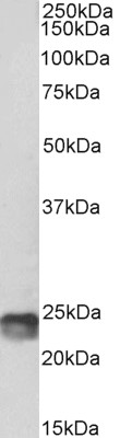
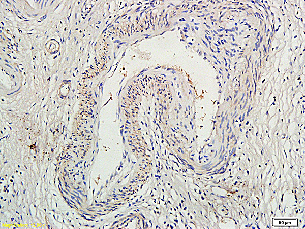
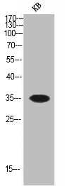
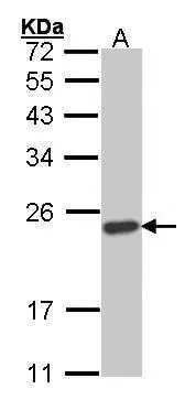
Anti-SOCS1 Antibody (Biotin)
A83760
ApplicationsWestern Blot, ELISA
Product group Antibodies
ReactivityHuman, Mouse
Overview
- SupplierAntibodies.com
- Product NameAnti-SOCS1 Antibody (Biotin)
- Delivery Days Customer7
- ApplicationsWestern Blot, ELISA
- CertificationResearch Use Only
- ClonalityPolyclonal
- Concentration500 ug/ml
- ConjugateBiotin
- HostGoat
- IsotypeIgG
- Scientific DescriptionGoat polyclonal antibody to SOCS1 (Biotin).
- ReactivityHuman, Mouse
- UNSPSC12352203
Related products
Product group Antibodies
Anti-SOCS1 Antibody144-07754
ApplicationsWestern Blot
ReactivityHuman, Mouse, Rat
TargetSOCS1
- SizePrice

Product group Antibodies
Anti-SOCS1 Antibody Picoband(r)A00448-CARRIER-FREE
ApplicationsWestern Blot, ImmunoHistoChemistry
ReactivityHuman, Mouse, Rat
TargetSOCS1
- SizePrice
Product group Antibodies
SOCS1 Polyclonal AntibodyBS-0113R
ApplicationsImmunoFluorescence, Western Blot, ELISA, ImmunoHistoChemistry, ImmunoHistoChemistry Frozen, ImmunoHistoChemistry Paraffin
ReactivityHuman, Mouse, Rat
TargetSOCS1
- SizePrice
Product group Antibodies
SOCS1 AntibodyCSB-PA004129
ApplicationsWestern Blot, ELISA
ReactivityHuman, Mouse, Rat
TargetSOCS1
- SizePrice
Product group Antibodies

Goat anti-SOCS1, biotinylatedEB05040-B
ApplicationsWestern Blot, ELISA
ReactivityBovine, Canine, Human, Mouse, Rat
TargetSOCS1
- SizePrice
Product group Antibodies
ApplicationsImmunoPrecipitation, Western Blot, ImmunoCytoChemistry, ImmunoHistoChemistry
ReactivityMouse, Porcine, Rat
TargetSOCS1
- SizePrice
Product group Antibodies

SOCS1 AntibodyLS-C402549
ApplicationsWestern Blot, ELISA, ImmunoHistoChemistry
ReactivityHuman, Mouse, Rat
TargetSOCS1
- SizePrice
Product group Antibodies
SOCS1 antibodyGTX100657
ApplicationsImmunoFluorescence, Western Blot, ImmunoCytoChemistry, ImmunoHistoChemistry, ImmunoHistoChemistry Paraffin
ReactivityHuman, Mouse
TargetSOCS1
- SizePrice

Product group Antibodies
Anti-SOCS1 AntibodyHPA074108
ApplicationsImmunoCytoChemistry
ReactivityHuman
TargetSOCS1
- SizePrice