


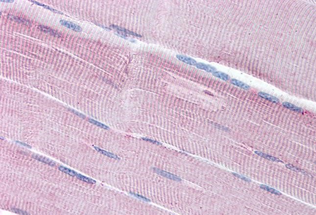
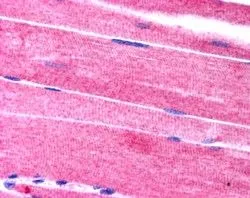
Immunohistochemical staining of human cerebral cortex, colon, lymph node and testis using Anti-SORBS1 antibody HPA027559 (A) shows similar protein distribution across tissues to independent antibody HPA043084 (B).

Immunohistochemical staining of human cerebral cortex, colon, lymph node and testis using Anti-SORBS1 antibody HPA027559 (A) shows similar protein distribution across tissues to independent antibody HPA043084 (B).
Anti-SORBS1 Antibody
HPA027559
ApplicationsWestern Blot, ImmunoCytoChemistry, ImmunoHistoChemistry
Product group Antibodies
ReactivityHuman
TargetSORBS1
Overview
- SupplierAtlas Antibodies
- Product NameAnti-SORBS1 Antibody
- Delivery Days Customer4
- ApplicationsWestern Blot, ImmunoCytoChemistry, ImmunoHistoChemistry
- CertificationResearch Use Only
- ClonalityPolyclonal
- ConjugateUnconjugated
- Gene ID10580
- Target nameSORBS1
- Target descriptionsorbin and SH3 domain containing 1
- Target synonymsCAP, FLAF2, R85FL, SH3D5, SH3P12, SORB1, sorbin and SH3 domain-containing protein 1, Fas-ligand associated factor 2, SH3 domain protein 5, c-Cbl associated protein, ponsin
- HostRabbit
- IsotypeIgG
- Protein IDQ9BX66
- Protein NameSorbin and SH3 domain-containing protein 1
- Scientific DescriptionRecombinant Protein Epitope Signature Tag (PrEST) antigen sequence
- ReactivityHuman
- Storage Instruction-20°C,2°C to 8°C
- UNSPSC41116161
Datasheet
MSDS
Related products
Product group Antibodies
Anti-SORBS1 AntibodyA285960
ApplicationsELISA, ImmunoHistoChemistry
ReactivityHuman
- SizePrice

Product group Antibodies
Anti-SORBS1 Picoband(r) AntibodyA04426-3-CARRIER-FREE
ApplicationsFlow Cytometry, Western Blot, ELISA, ImmunoHistoChemistry
ReactivityHuman
TargetSORBS1
- SizePrice
Product group Antibodies

ApplicationsELISA, ImmunoHistoChemistry
ReactivityHuman, Mouse, Porcine, Rat
TargetSORBS1
- SizePrice
Product group Antibodies
SORBS1 antibody, C-termGTX24551
ApplicationsImmunoHistoChemistry, ImmunoHistoChemistry Paraffin
ReactivityHuman
TargetSORBS1
- SizePrice

Product group Antibodies
Anti-SORBS1 AntibodyHPA043084
ApplicationsWestern Blot, ImmunoHistoChemistry
ReactivityHuman
TargetSORBS1
- SizePrice
Product group Antibodies

SORBS1 / Ponsin AntibodyLS-C753817
ApplicationsWestern Blot, ELISA
ReactivityHuman
TargetSORBS1
- SizePrice