

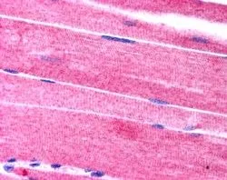
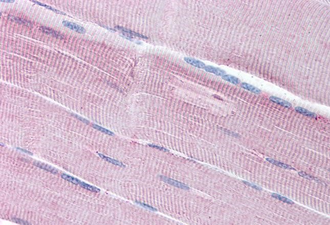
IHC-P analysis of human skeletal muscle using GTX24551 SORBS1 antibody, C-term. Antigen retrieval : citrate buffer Ph 6 Dilution : 2.5μg/ml
IHC-P analysis of human skeletal muscle using GTX24551 SORBS1 antibody, C-term. Antigen retrieval : citrate buffer Ph 6 Dilution : 2.5μg/ml
SORBS1 antibody, C-term
GTX24551
ApplicationsImmunoHistoChemistry, ImmunoHistoChemistry Paraffin
Product group Antibodies
ReactivityHuman
TargetSORBS1
Overview
- SupplierGeneTex
- Product NameSORBS1 antibody, C-term
- Delivery Days Customer9
- Application Supplier NoteIHC-P: 2.5microg/ml. *Optimal dilutions/concentrations should be determined by the researcher.Not tested in other applications.
- ApplicationsImmunoHistoChemistry, ImmunoHistoChemistry Paraffin
- CertificationResearch Use Only
- ClonalityPolyclonal
- Concentration0.50 mg/ml
- ConjugateUnconjugated
- Gene ID10580
- Target nameSORBS1
- Target descriptionsorbin and SH3 domain containing 1
- Target synonymsCAP, FLAF2, R85FL, SH3D5, SH3P12, SORB1, sorbin and SH3 domain-containing protein 1, Fas-ligand associated factor 2, SH3 domain protein 5, c-Cbl associated protein, ponsin
- HostGoat
- IsotypeIgG
- Protein IDQ9BX66
- Protein NameSorbin and SH3 domain-containing protein 1
- Scientific DescriptionThis gene encodes a CBL-associated protein which functions in the signaling and stimulation of insulin. Mutations in this gene may be associated with human disorders of insulin resistance. Alternative splicing results in multiple transcript variants. [provided by RefSeq, Mar 2014]
- ReactivityHuman
- Storage Instruction-20°C or -80°C,2°C to 8°C
- UNSPSC41116161
Datasheet
Related products
Product group Antibodies
Anti-SORBS1 AntibodyA285960
ApplicationsELISA, ImmunoHistoChemistry
ReactivityHuman
- SizePrice

Product group Antibodies
Anti-SORBS1 Picoband(r) AntibodyA04426-3-CARRIER-FREE
ApplicationsFlow Cytometry, Western Blot, ELISA, ImmunoHistoChemistry
ReactivityHuman
TargetSORBS1
- SizePrice
Product group Antibodies

ApplicationsELISA, ImmunoHistoChemistry
ReactivityHuman, Mouse, Porcine, Rat
TargetSORBS1
- SizePrice

Product group Antibodies
Anti-SORBS1 AntibodyHPA027559
ApplicationsWestern Blot, ImmunoCytoChemistry, ImmunoHistoChemistry
ReactivityHuman
TargetSORBS1
- SizePrice
Product group Antibodies

SORBS1 / Ponsin AntibodyLS-C753817
ApplicationsWestern Blot, ELISA
ReactivityHuman
TargetSORBS1
- SizePrice