

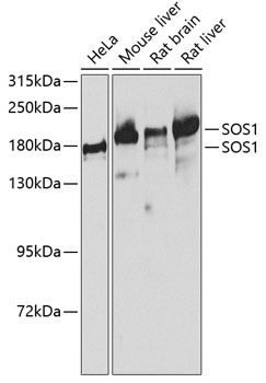
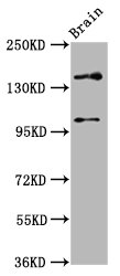
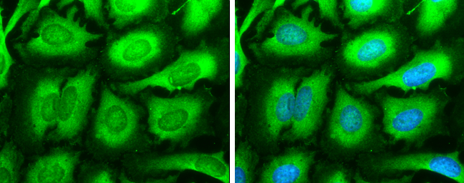
Anti-SOS1 Antibody
A14419
ApplicationsWestern Blot
Product group Antibodies
ReactivityHuman, Mouse, Rat
Overview
- SupplierAntibodies.com
- Product NameAnti-SOS1 Antibody
- Delivery Days Customer7
- ApplicationsWestern Blot
- CertificationResearch Use Only
- ClonalityPolyclonal
- ConjugateUnconjugated
- HostRabbit
- IsotypeIgG
- Scientific DescriptionRabbit polyclonal antibody to SOS1.
- ReactivityHuman, Mouse, Rat
- UNSPSC12352203
Related products

Product group Antibodies
Anti-SOS1 Antibody Picoband(r)A00837-1-CARRIER-FREE
ApplicationsFlow Cytometry, Western Blot, ELISA, ImmunoHistoChemistry
ReactivityHuman, Mouse, Rat
TargetSOS1
- SizePrice
Product group Antibodies
Anti-SOS1 Antibody144-03272
ApplicationsImmunoFluorescence, Western Blot
ReactivityHuman, Mouse, Rat
TargetSOS1
- SizePrice
Product group Antibodies
SOS1 Recombinant Antibody, Biotin ConjugatedBSM-61476R-BIOTIN
ApplicationsWestern Blot, ImmunoHistoChemistry, ImmunoHistoChemistry Frozen, ImmunoHistoChemistry Paraffin
ReactivityHuman, Mouse, Rat
TargetSOS1
- SizePrice
Product group Antibodies
SOS1 AntibodyCSB-PA726788LA01HU
ApplicationsImmunoFluorescence, Western Blot, ELISA, ImmunoHistoChemistry
ReactivityHuman, Rat
TargetSOS1
- SizePrice
Product group Antibodies
Sos1 Polyclonal AntibodyCAC11454
ApplicationsImmunoFluorescence, Western Blot, ELISA, ImmunoHistoChemistry
ReactivityRat
TargetSOS1
- SizePrice
Product group Antibodies

SOS1 AntibodyLS-C408689
ApplicationsWestern Blot
ReactivityHuman, Mouse, Rat
TargetSOS1
- SizePrice
Product group Antibodies
SOS1 antibodyGTX133774
ApplicationsImmunoFluorescence, Western Blot, ImmunoCytoChemistry
ReactivityHuman
TargetSOS1
- SizePrice
Product group Antibodies
Anti-SOS1 AntibodyCAB3272
ApplicationsWestern Blot, ELISA
ReactivityHuman
TargetSOS1
- SizePrice