


Anti-SPAG16 Antibody
A82659
ApplicationsFlow Cytometry, ImmunoFluorescence, ELISA, ImmunoHistoChemistry
Product group Antibodies
ReactivityHuman, Mouse
Overview
- SupplierAntibodies.com
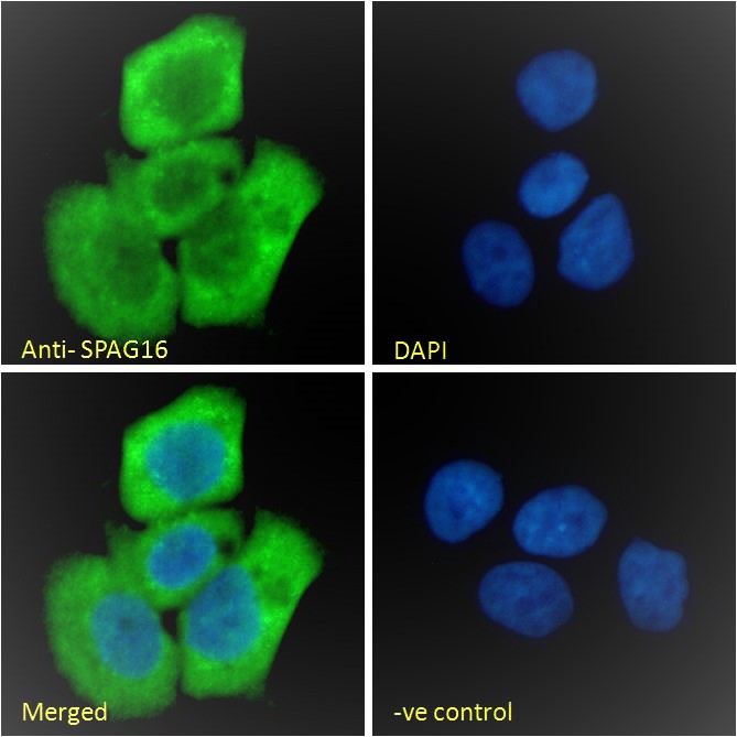
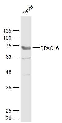
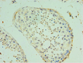
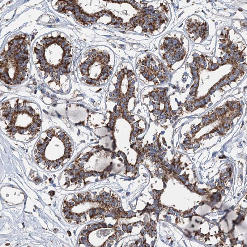
- Product NameAnti-SPAG16 Antibody
- Delivery Days Customer7
- ApplicationsFlow Cytometry, ImmunoFluorescence, ELISA, ImmunoHistoChemistry
- CertificationResearch Use Only
- ClonalityPolyclonal
- Concentration500 ug/ml
- ConjugateUnconjugated
- HostGoat
- IsotypeIgG
- Scientific DescriptionGoat polyclonal antibody to SPAG16.
- ReactivityHuman, Mouse
- UNSPSC12352203
Related products

Product group Antibodies
Anti-SPAG16 Antibody Picoband(r)A09080-3-CARRIER-FREE
ApplicationsFlow Cytometry, Western Blot, ELISA
ReactivityHuman, Mouse, Rat
TargetSPAG16
- SizePrice
Product group Antibodies
SPAG16 Polyclonal AntibodyBS-11485R
ApplicationsImmunoFluorescence, Western Blot, ELISA, ImmunoCytoChemistry, ImmunoHistoChemistry, ImmunoHistoChemistry Frozen, ImmunoHistoChemistry Paraffin
ReactivityBovine, Canine, Equine, Human, Mouse, Rabbit, Rat, Sheep
TargetSPAG16
- SizePrice
Product group Antibodies
References

Goat anti-SPAG16 (aa139-152)EB09826
ApplicationsFlow Cytometry, ImmunoFluorescence, ELISA, ImmunoHistoChemistry
ReactivityBovine, Canine, Human, Mouse
TargetSPAG16
- SizePrice
Product group Antibodies
SPAG16 AntibodyCSB-PA818674ESR1HU
ApplicationsELISA, ImmunoHistoChemistry
ReactivityHuman
TargetSPAG16
- SizePrice
Product group Antibodies

SPAG16 AntibodyLS-C349176
ApplicationsWestern Blot, ImmunoHistoChemistry
ReactivityHuman, Mouse
TargetSPAG16
- SizePrice
Product group Antibodies
Anti-SPAG16 AntibodyHPA037542
ApplicationsWestern Blot, ImmunoCytoChemistry, ImmunoHistoChemistry
ReactivityHuman
TargetSPAG16
- SizePrice